
Dear Wei, This is a visualization issue (rather than a processing issue). Open your T2 first, and then your T1 on top, and there shouldn’t be any black blocks. Cheers, /Eugenio
Juan Eugenio Iglesias Senior research fellow CMIC (UCL), MGH (HMS) and CSAIL (MIT) http://www.jeiglesias.com
From: freesurfer-bounces@nmr.mgh.harvard.edu on behalf of Wei Shao wshao@research.baycrest.org Reply-To: Freesurfer support list freesurfer@nmr.mgh.harvard.edu Date: Tuesday, February 23, 2021 at 16:49 To: Freesurfer support list freesurfer@nmr.mgh.harvard.edu Subject: [Freesurfer] Hippocampus subfiled segmantation addtional scan
External Email - Use Caution Hi, Freesurfer experts
I used the Freesurfer7.1.0 version for the hippocampus subfield segmentation, and I use a coronal T2 scan as an additional to do the segmentation .
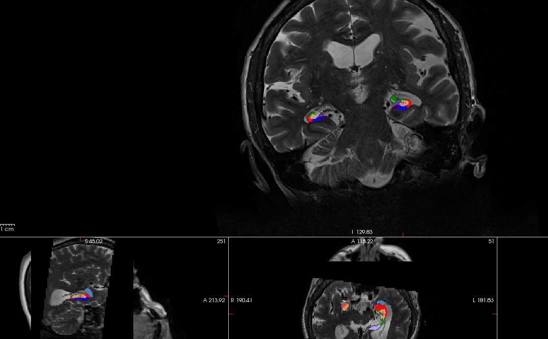
[cid:image001.png@01D70A04.62CE8E90]
I'm wondering that the reason for the two black blocks in the sagittal and axial view is because I only use the coronal T2 anat scan for the additional scan?
Can I add another axial scan for the T2 subfield segmentation without deleting anything (like hippocampal-subfields-T2.T2.log)? Thanks
Best, Wei