
Hello,
I am interested in the cortical parcellations at the pial (grey matter) and white matter layers
I have extracted grey matter (pial) and white matter (white) labels for a gyrus (right superior frontal gyrus, label 2028)
mri_annotation2label --subject s1/12copy --hemi rh --labelbase ./12copy/label/rh.cortex --surface pial
mri_annotation2label --subject s1/12copy --hemi rh --labelbase ./12copy/label/rh.cortex --surface white
then converted the above 2 files (renamed to ...-wm and ...-gm) for the superior frontal gyrus to nifti's for viewing in other applications
mri_label2vol --label ./label/rh.cortex-028.label --o rh-cortex-028-rsfg-wm.nii --identity --temp ./mri/T1.mgz
mri_label2vol --label ./label/rh.cortex-028.label --o rh-cortex-028-rsfg-gm.nii --identity --temp ./mri/T1.mgz
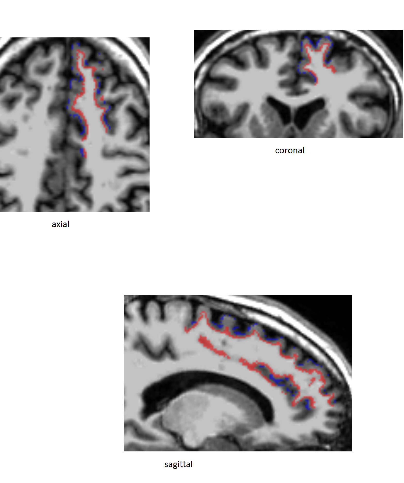
My goal is to run some statistics on 2 nifti's, which requires continuity in the nonzero voxels, in at least 1 of the 3 directions (x/y/z). This isn't the case for this gyri, particularly at the pial layer (shown in blue in the attachment). Is there a way in which this could be achieved? Have I missed any steps?
Many thanks in advance.
Shads
The University of Edinburgh is a charitable body, registered in Scotland, with registration number SC005336.