
External Email - Use Caution
Hi Anastasia,
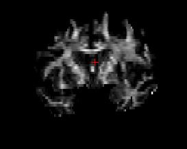
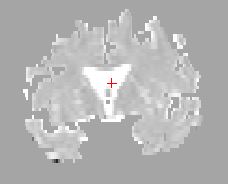
I tried to visualize the RD.nii with the following steps, however, it doesn't represent well as fa.nii does. (attached below).
1) RD = (L2+L3)/2: fslmaths dtifit_L2.nii -add dtifit_L3 -div 2 dtifit_RD.nii 2) mri_vol2vol --mov dmri/lowb.nii --targ mri/wmparc.mgz --inv --interp nearest --o mri/wmparc2diff.mgz --reg dmri/xfms/anatorig2diff.bbr.dat --no-save-reg 3) mri_mask dmri/dtifit_RD.nii mri/wmparc2diff.mgz dmri/rd-masked.mgz 4) freeview -v dmri/dtifit_RD.nii dmri/rd-masked.mgz
If I tried to color-code every diffusivity (rd, ad, fa), I failed by adding "edgecolor=red(blue, or yellow)" following, what are the other options?
freeview -v
dmri/dtifit_FA.nii:reg=dmri/xfms/anatorig2diff.bbr.dat:edgecolor=red dmri/dtifit_AD.nii:reg=dmri/xfms/anatorig2diff.bbr.dat:edgecolor=yellow...
FA.nii RD.nii [image: FA2.JPG] [image: RD1.JPG]
Thank you! Best, Jackie
On Mon, Nov 22, 2021 at 7:27 PM Yendiki, Anastasia AYENDIKI@mgh.harvard.edu wrote:
See "viewing volumes with freeview" in the tutorial: https://secure-web.cisco.com/10RdLE2TlyCn6SHIX3mLEiv_Rt4eMDoWoVQI8x2WCLgHYOa...
When you view the aseg, aparc+aseg, or any other FreeSurfer segmentation volume, make sure that the colormap is not grayscale but "look-up table".
You can get the AD and RD from the other outputs: AD = L1 RD = (L2+L3)/2
*From:* freesurfer-bounces@nmr.mgh.harvard.edu < freesurfer-bounces@nmr.mgh.harvard.edu> on behalf of Zeng, Qi < qi.zeng@icahn.mssm.edu> *Sent:* Monday, November 22, 2021 5:24 PM *To:* Freesurfer support list freesurfer@nmr.mgh.harvard.edu *Subject:* [Freesurfer] Freeview grey matter, white matter and Tracula outputs
External Email - Use CautionHi,
If I want to freeview fully segmented grey matter, white matter, and Tracula outputs, are the following commands correct to the request?
# to visualize the segmented cortical outputs freeview -v mri/aparc+aseg.mgz Question: why is one hemisphere brighter than the other? How can I adjust so that they are equally bright?
# to visualize the segmented subcortical outputs freeview -v mri/aseg.mgz Question: why is only one hemisphere showing and how to add the other one?
$ to visualize AD. RD, FA and color-coded each freeview -v dmri/dtifit_FA.nii Question: I cannot find the nifti outputs of AD and RD (however, the stats generated). where can I find it? Is there an example of how to color-code them for freeview?
Thank you! Best, Jackie _______________________________________________ Freesurfer mailing list Freesurfer@nmr.mgh.harvard.edu https://secure-web.cisco.com/1wqxCmEbMWS5bXmDde-Cm4n5E1m4dp1UJY3EPioSjfVhLvt... The information in this e-mail is intended only for the person to whom it is addressed. If you believe this e-mail was sent to you in error and the e-mail contains patient information, please contact the Mass General Brigham Compliance HelpLine at http://secure-web.cisco.com/1ZwvmXrSa5vi0hTZk0ZMYKm8seFVH4b6-ZuTUhJtG_OgzVU0... . If the e-mail was sent to you in error but does not contain patient information, please contact the sender and properly dispose of the e-mail. Please note that this e-mail is not secure (encrypted). If you do not wish to continue communication over unencrypted e-mail, please notify the sender of this message immediately. Continuing to send or respond to e-mail after receiving this message means you understand and accept this risk and wish to continue to communicate over unencrypted e-mail.