
External Email - Use Caution
Hi Douglas,
Did you have time to look at my new questions ?
Best, Matthieu
Le ven. 30 nov. 2018 à 12:15, Matthieu Vanhoutte < matthieuvanhoutte@gmail.com> a écrit :
Hi Douglas,
Thank you for answering. Please find below new questions.
Le ven. 30 nov. 2018 à 00:00, Greve, Douglas N.,Ph.D. < DGREVE@mgh.harvard.edu> a écrit :
Hi Matthieu, sorry for the delay
On 11/29/2018 12:50 PM, Matthieu Vanhoutte wrote:
External Email - Use CautionDear Freesurfer's experts,
I tried to use PETSurfer to correct partial volume effect on my FDG PET
images, testing both Muller-Gartner and RBV corrections.
I ran the commands specified in PETSurfer website and used the two
following commands for both MGX and RBV corrections respectively:
mri_gtmpvc --i PET.nii.gz --reg register.dof6.lta --psf-col 5.51
--psf-row 5.51 --psf-slice 5.9 --seg gtmseg.mgz --default-seg-merge --auto-mask PSF .01 --mgx .01 --o ./gtmpvc.output
mri_gtmpvc --i PET.nii.gz --reg register.dof6.lta --psf-col 5.51
--psf-row 5.51 --psf-slice 5.9 --seg gtmseg.mgz --default-seg-merge --auto-mask PSF .01 --rbv --o rbv.output.orig
- However, I found that cortical output mgx.ctxgm.nii.gz of MGX
correction encompass more than just GM and values at the boundaries of mgx.ctxgm.nii.gz seem to me very high or aberrant. This is expected. The MG method gives you a value every place that there is GM signal *in the PET volume after partial volume effects*. So basically, if you were to take the cortical ribbon and smooth it by your PSF, every non-zero voxel has some GM in it (which is why the edges are so high). When you run it with --mgx .01, it will exclude voxels that have less than 1% GM after smoothing. If you you are disturbed by the wide ribbon, just make the threshold higher. In theory, every point along the surface normal gives you a valid answer, but the further from the center of the ribbon, the noisier it is going to be, so we generally only sample it at the center (--projfrac 0.5 to mri_vol2surf).
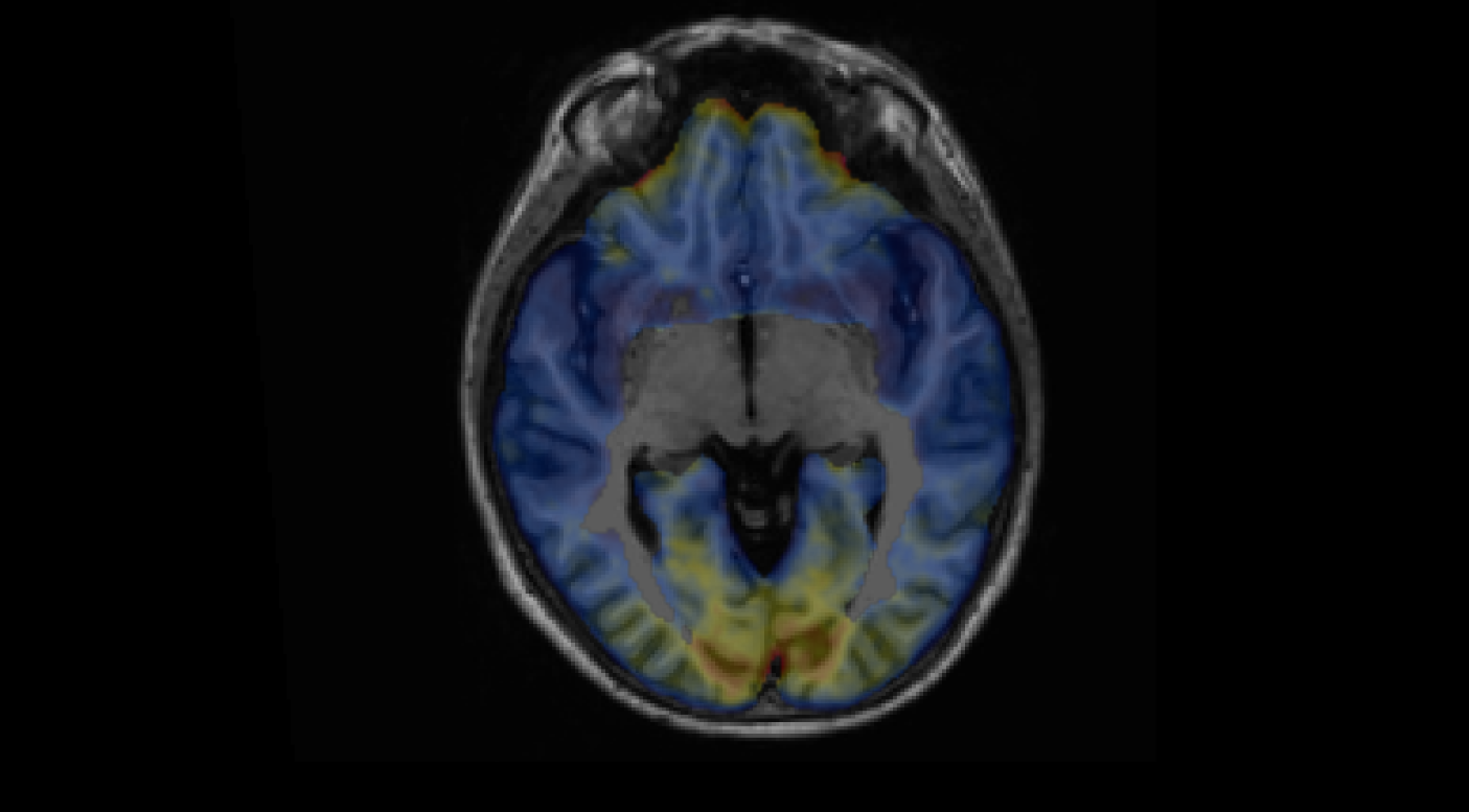
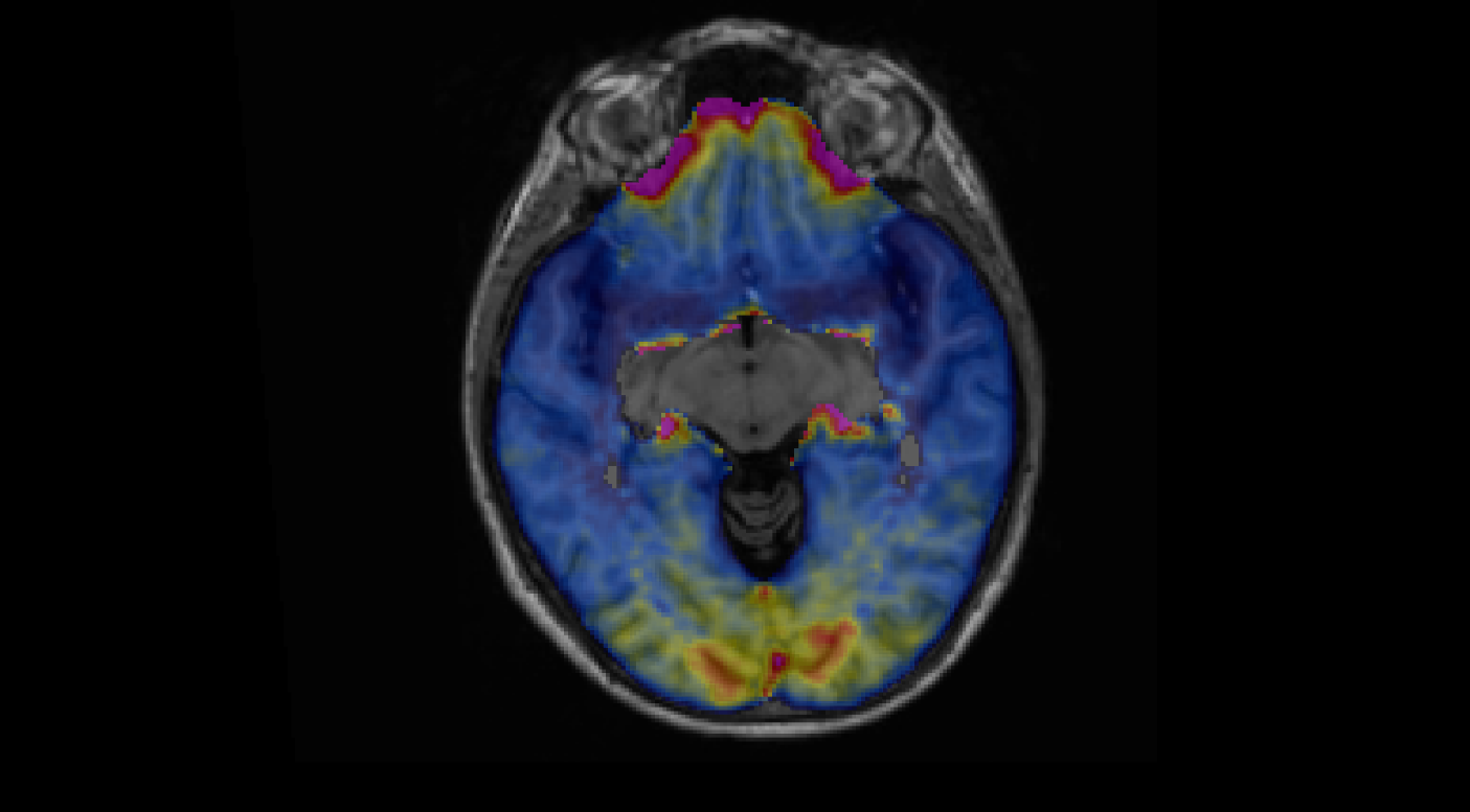
Basically, please find below the mgx.ctxgm with threshold set at 0.01: [image: image.png]
Then threshold set at 0.1: [image: image.png]
Values at some parts of the cortex (olfactory, visual) are not the same between the two thresholds. In the first one in these parts of the brain, values are higher than the second and seem kind of aberrant. Is there no reason to prefer a threshold at 0.1 than 0.01 ? For example, in (Douglas et al., 2016, NeuroImage) a threshold of 0.3 has been found to be optimal: how determine visually or quantitatively this optimal threshold ?
- Concerning RBV correction, output rbv.nii.gz seems to me following
more precisely the GM ribbon. However contrary to what is said in PETSurfer website, rbv.nii.gz seems to be in the anatomical space (not in native PET) at the resolution of gtmseg.mgz. How then map rbv.nii.gz to the anatomical space when mapping the volume to the surface ? Where does it say this? It should be in the anatomical space in the sense that it shares an RAS space with the conformed volume (aseg does gtmseg.mgz). This means that you can use --regheader with mri_vol2surf or mri_vol2vol when mapping into another space.
In https://surfer.nmr.mgh.harvard.edu/fswiki/PetSurfer it says that "mgx.ctxgm is in same resolution of the input PET", which is the case since resolution and orientation are identical to native PET. The PETsurfer tutorial then explains that "bbpet2anat.lta. is a registration file that can be used to map the output PET volume (in the mask bounding box) to the anatomical space".
However, when I open rbv.nii file it is not in native PET resolution and orientation but those of gtmseg.mgz (anatomical space but with resolution of 0.5x0.5x05 mm). Why these differences between these two methods of PVC and which registration file then to use when mapping rbv.nii to the surface (rbv2anat.lta ?) ? I think I can't use directly --regheader since resolution of rbv.nii is 0.5 mm3 whereas anatomical space is of 1 mm3.
- What are the advantages/inconveniences of RBV vs GMX ?
Not entirely sure. RBV may be more precise since it at least has the ability to correct for the PVE across the bank of a sulcus, but the two banks have to be in different ROIs. The bad news is that the RBV correction depends on the ROIs that you use.
MGX doesn't correct PVE across the bank of a sulcus ?
By saying that "RBV correction depends on the ROIs that you use", do you mean the parcellation (aparc or aparc.a2009s) you give to the gtmseg command ? If this is the case is there a better compromise ?
- Would it be beneficial to upsample native PET to the anatomical
resolution before launching gtmpvc in order to preserve the high resolution of the anatomical tissues during partial volume correction ? No, this is all taken care of in mri_gtmpvc.
Could you have a look at and give me back your opinion on these
questions ? I could send the associated files if needed.
Thank you.
Best, Matthieu
Freesurfer mailing list Freesurfer@nmr.mgh.harvard.edu https://mail.nmr.mgh.harvard.edu/mailman/listinfo/freesurfer
Freesurfer mailing list Freesurfer@nmr.mgh.harvard.edu https://mail.nmr.mgh.harvard.edu/mailman/listinfo/freesurfer