
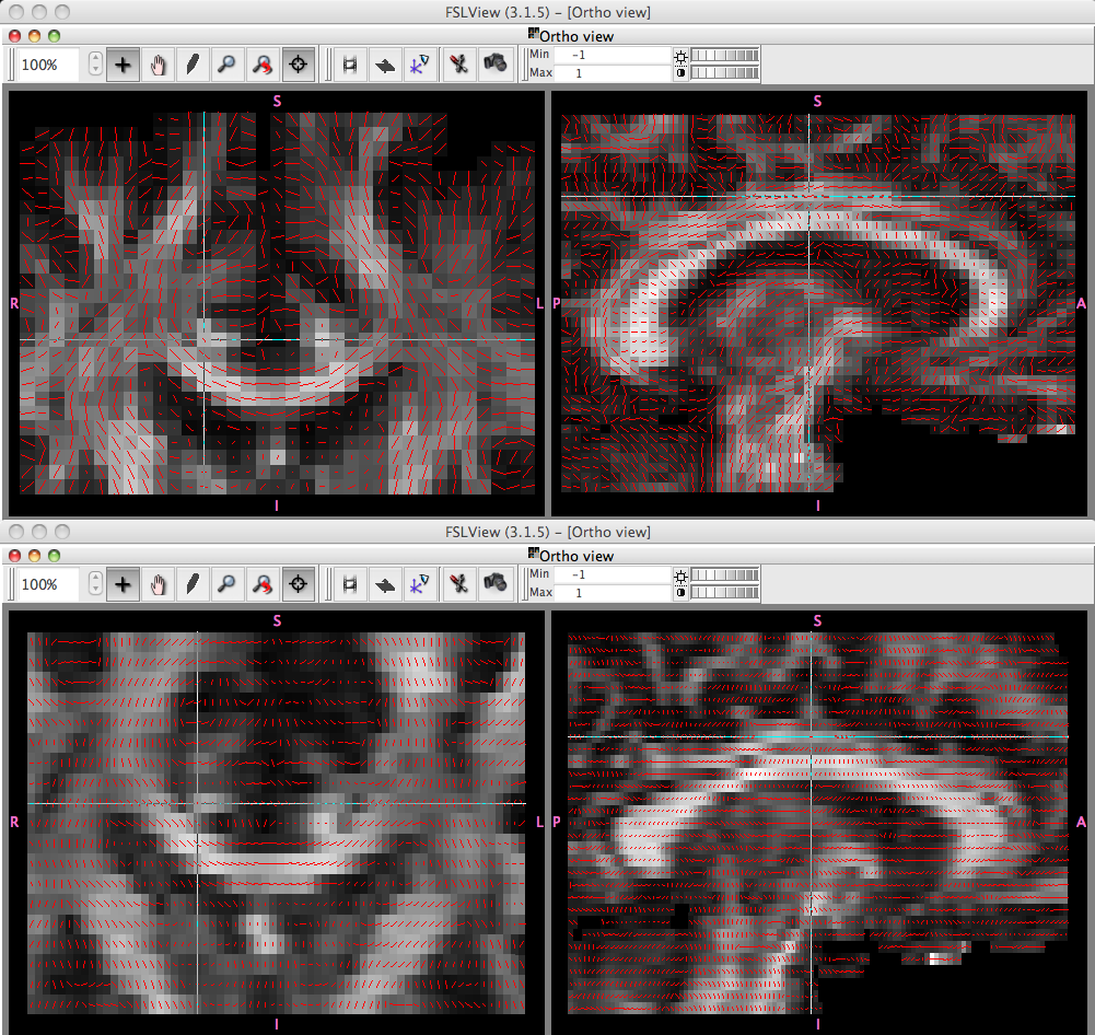
Hi Ansgar - I'm attaching a screenshot of dtifit outputs, FA and primary eigenvector directions. The top is one of our own subjects, the bottom is your subject where tracula failed. You can see that the directions in your subject in the sagittal view are not following the cingulum. This is probably b/c of bvecs not having the right direction.
If the tensor fit isn't right, then the bedpostx ball-and-stick fit (that trac-all uses) won't be right either. So it's a good troubleshooting tool.
Also, if you have the option, I'd switch to an isotropic resolution for your DWI acquisition. Anisotropic voxels are generally considered to introduce bias in diffusion analysis - not sure to what extent this will hurt you but it's not going to help you to have 1mm in-plane and 2mm slice thickness. I'd switch to 2mm isotropic, it'd help your SNR and avoid the direction bias.
But for the existing data, let's fix the bvecs first and see.
Hope this helps, a.y
On Thu, 15 Mar 2012, Ansgar Furst wrote:
Dear All, Please ignore my earlier incomplete message with the same title! We are using the ADNI Acc 3D FSPGR for structurals and a 30 directional (5 B0s) dual-spin repeat sequence for DTI (on a 3T GE 750 discovery) which so far rendered really nice results. However, recently I encountered data from several healthy young subjects where the TRACULA fiber tract estimations would suggest severely "crippled" tracts which, however, is not consistent with their FA maps. The crippling is most visible when comparing left vs right cingulate gyri. In some cases either the left or right side is almost completely gone. Has anyone else experienced similar issues and found a remedy? Thanks a lot, Ansgar