
External Email - Use Caution
Hi Freesurfers,
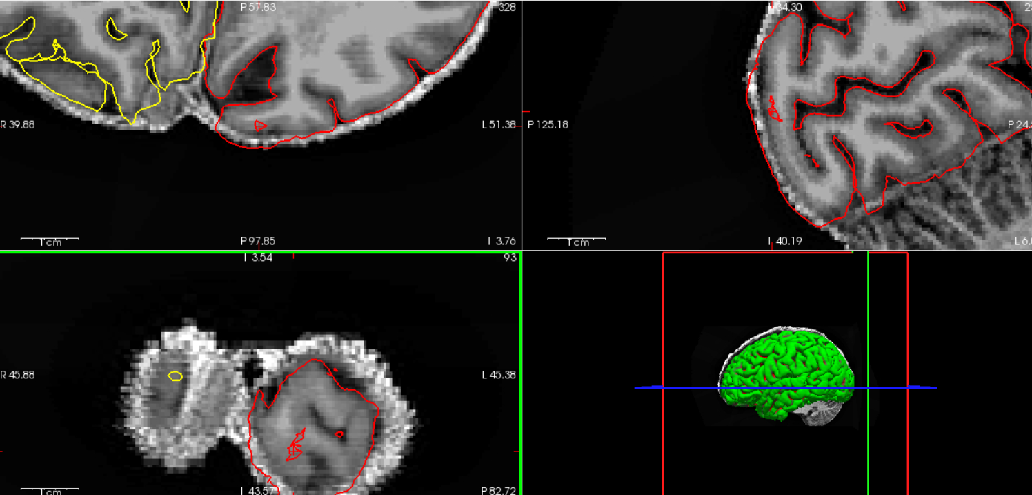
I have a question reagrding to how the pial surface is generated. For an oversimplified description, I assume each subject's pial surface was initially created with a spherical shaped 3D-topology model. Then each verticie on the sphere is assigned to specific location of the pial surface to cover the subject's brain. If that is the case, then I don't understand the result illustrated below in the figure. The figure is showing anatomical T1w image overlay with left and right pial surface from recon-all (command used at the end), while the red cross is targeting a pial surface located within cortical region. Although there is some mis-segmentation error nearby, I don't see the pial surface inside of the cortical region is connecting to any outer surface when I move the image slice back and forth. Is this allowed or even possible if the initial spherical surface can generate such discrete surface that is disconnected from the whole outer brain region?
[cid:20a64222-bc84-4c74-9935-ffd295eac833]
Does anyone know what is the cause of this and how to trouble shoot? Thanks in advance!
1. Freesurfer version: freesurfer-linux-ubuntu22_x86_64-7.4.1-20230614-7eb8460 2. Platform: Ubuntu 24.04.2 LTS 3. uname -a: Linux hawaii 6.11.0-26-generic #26~24.04.1-Ubuntu SMP PREEMPT_DYNAMIC Thu Apr 17 19:20:47 UTC 2 x86_64 x86_64 x86_64 GNU/Linux 4. Command and steps used for recon-all: * recon-all -autorecon1 -s <subj_id> -i <mp2rage_input> * check and verify skull strip quality * recon-all -autorecon2 -autorecon3 -s <subj_id>
Best,
Yujia Huang
Research Engineer
Advanced Imaging Research Center
UT Southwestern Medical Center
2201 Inwood Rd.
Dallas, TX, USA, 75390
Office: 214.645.5241
Pronouns: He, Him, His
________________________________
UT Southwestern
Medical Center
The future of medicine, today.