
Hi Martin,
Here are the instructions: https://surfer.nmr.mgh.harvard.edu/fswiki/FtpFileExchange
If you can, please upload both T1, T2, and FLAIR scans of that particular subject.
Thanks, Stefano ________________________________ Da: Martin Kavec martin.kavec@gmail.com Inviato: martedì 16 agosto 2022 03:42 A: Cerri, Stefano SCERRI@mgh.harvard.edu Cc: freesurfer@nmr.mgh.harvard.edu freesurfer@nmr.mgh.harvard.edu Oggetto: Re: [Freesurfer] Large number of false negative MS lesions in SAMSEG
External Email - Use Caution
Hi Stefano,
thanks a lot for advice on t2 intensity pattern. I ran couple of cases and I clearly see an improvement. I do not need to reduce threshold at all to get better results. Although the number of FN dropped by roughly 1/2, there are still quite a lot of them. For example, the lesion which I sent in my first email is still left behind. I am happy to send you that particular case. May I ask you for instructions, where to upload it?
Thanks,
Martin
On Sun, Aug 14, 2022 at 10:52 PM Cerri, Stefano <SCERRI@mgh.harvard.edumailto:SCERRI@mgh.harvard.edu> wrote: Hi Martin,
Can you try to remove the lesion intensity constraint on the T2 data (i.e., by modifying your command with --lesion-mask-pattern 0 1 0) for a couple of cases and let us know if it helps?
If not, would you be able to share with us one case where it fails?
FYI: you can save a lesion probability map directly by using the flag "--save-posteriors Lesions" so you don't need to try different thresholds.
Thanks, Stefano
Subject: [Freesurfer] Large number of false negative MS lesions in SAMSEG Date: Sun, 14 Aug 2022 20:54:50 +0200 From: Martin Kavec martin.kavec@gmail.commailto:martin.kavec@gmail.com Reply-To: Freesurfer support list freesurfer@nmr.mgh.harvard.edumailto:freesurfer@nmr.mgh.harvard.edu To: Freesurfer mailing list freesurfer@nmr.mgh.harvard.edumailto:freesurfer@nmr.mgh.harvard.edu
External Email - Use Caution
Hello,
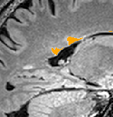
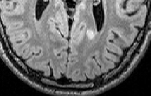
I am testing SAMSEG on my group of MS patients. Unfortunately, even when dropping threshold to 0.005 I have large number of false negative lesions left behind by SAMSEG (80% in about 75 cases I tried), which are clearly visible lesions. I ran/checked registration between inputs, which is typically very good to excellent. Images are all 3D acquisitions , mprage, flair, and t2 with ~1 mm3 isotropic voxel. Here is the command, which I ran:
run_samseg --input mprage_reg.nii.gz flair.nii.gz t2_reg.nii.gz --pallidum-separate --lesion --lesion-mask-pattern 0 1 1 --threshold 0.005 --output samseg_threshold_0.005
and example of the lesion in occipital white matter in sagittal and axial orientations.
[image.png] [image.png]
I would very much appreciate any leads as to how to improve the segmentation.
Thanks,
Martin
The information in this e-mail is intended only for the person to whom it is addressed. If you believe this e-mail was sent to you in error and the e-mail contains patient information, please contact the Mass General Brigham Compliance HelpLine at https://www.massgeneralbrigham.org/compliancelinehttps://secure-web.cisco.com/1qmbHsOu4BhgJgl2jzOYTijx2JsTOaHP8LBIv1cqbkI9kd2nDxMe09S7s726qz6okDyvByRILwRyIrX3jvD39UBA2-7bN7cb1AhGmdxTjhb5MXmPEIWhxlv9JU2pvPbvw_aE1pKfhqQmP0gjXIej7Diqz1znOkjVvJBUTpJpUoscIvubovZ8nzzED7zMgwWEr3EABQIqiuy1rGiY5JEak6_BY-1j_5EdmKlEwkbr6uhlb56F1o3aFTF8gM1bD0kW1386b-Z2cJM3EiJ5YehKRxGZcQxofmNMXiCqh7N0ZbscWraCzkQCcKhpTR-Bf8Zcg/https%3A%2F%2Fwww.massgeneralbrigham.org%2Fcomplianceline .