
Dear Sebastian,
Attached is the *.orig for both recon (green being the one with wm error and red being the one with error). Unfortunately they are not identical; a section of the dura is misclassified as wm in the red delineation. As mentionned before, both orig.mgz are numerically identical in that area. As I also mentionned before, the recon were ran on the same machine, same os, same binaries.
You expressed some confusion as to what were trying to achieve. We want to figure out why, given numerically almost identical inputs, Freesurfer correctly delineates wm in one case but fails in the other. Unfortunately this type of error appears to be systematic in some of our data.
Vincent.
On 26.08.2014 16:22, Sebastian Moeller wrote:
Hi Vincent hi
Melanie,
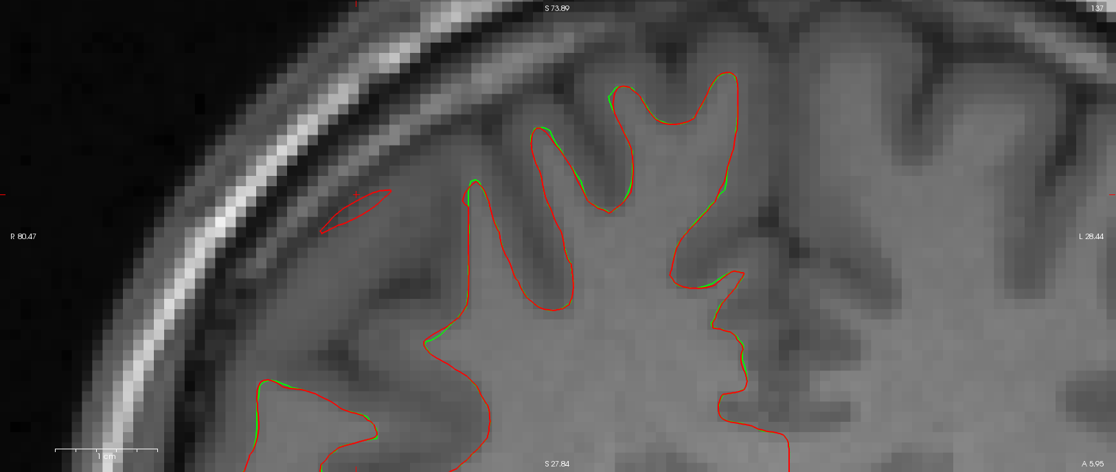
please show us the *.orig surfaces on the critical slice. I
predict that these will be identical, If so you can ignore everything except brain.finalsurfs.mgz. Are the two versions of this file identical around the area of mis-classified tissue? I predict that there will be a slight difference. Also if you take Vincent's brain.finalsurf.mgz and put this into Melanie's recon and redo the generation of the wm surfaces I predict that now Melinie's version will show the same problem.
On Aug 26, 2014, at 16:08 , Melanie Ganz <Melanie.Ganz@nru.dk [3]> wrote:
Hi Sebastian,
I'm afraid that the problem is
slightly more complicated. The inputs to recon-all are almost numerically identical (to the 4th decimal); when converted to orig.mgz, all voxels which are not identical (about only 700 of all the 1677216 voxels) vary only by 1 due to rounding. Their distribution is sparse and do not overlap with the white matter errors. Can such a small source of variation give this kind of difference? Ok, Freesurfer uses some random initialization, however as far as I know the seed is not random and set to 1234. We have looked at all the outputs created before wm.mgz (orig,nu,T1,brainmask,norm,nu_noneck,aseg,brain,wm,filled) and the only one showing a differences overlapping the wm errors is filled.mgz;
I think the filled.mgz is changed during the generation of the wm surfaces (at least temporarily)...
in all the other images the
differences are minor.
Are you running the analyses on exactly the
same machine and get different results or are you using two "identical" computers? I ask as I think there is a paper about the reproducibility of freesurfer reconstructions (http://www.plosone.org/article/info%3Adoi%2F10.1371%2Fjournal.pone.0038234 [4]) with different freesurfer versions or different hardware/operating systems that might explain a bit of systematic variance.
But I guess
I still do not exactly understand what the problem is that you want to solve here ….
Best Regards Sebastian
Thanks.
Vincent.
P.S.: I keep sen
stian Moeller wrote:
Hi Melanie,
I guess your post got me confused then, since you did not really
explain what your figure shows. With the information from this post I assume that red and yellow are wm and pia respectively from and recon and blue and green are from the second.
On Aug 26, 2014, at
15:16 , Melanie Ganz <Melanie.Ganz@nru.dk [1]> wrote:
Hi
Sebastian,
it is clear to us how to fix this, this is not the
issue here.
The issue is that my colleague and I ran the same
image, he used the original dicom as source and I a nifti version prepared for other processing,
Plain conversion of dicim to
nifty or further processing?
-- Melanie Ganz, MSc, Ph.D.
Neurobiology Research Unit
University of Copenhagen
Rigshospitalet
Rockefeller Center Juliane Maries Vej 28/30, 3.
DK-2100 Copenhagen Denmark phone: +45 3545 6718 e-mail:
Melanie.Ganz@nru.dk [2]
_______________________________________________
Freesurfer mailing
list
Freesurfer@nmr.mgh.harvard.edu
https://mail.nmr.mgh.harvard.edu/mailman/listinfo/freesurfer
The
information in this e-mail is intended only for the person to whom it is
addressed. If you believe this e-mail was sent to you in error and
the e-mail
contains patient information, please contact the Partners
Compliance HelpLine at
the e-mail was sent to you in error
but does not contain patient
information, please contact the sender and properly
dispose of the
e-mail.
Links: ------ [1] mailto:Melanie.Ganz@nru.dk [2] mailto:Melanie.Ganz@nru.dk [3] mailto:Melanie.Ganz@nru.dk [4] http://www.plosone.org/article/info%3Adoi%2F10.1371%2Fjournal.pone.0038234