
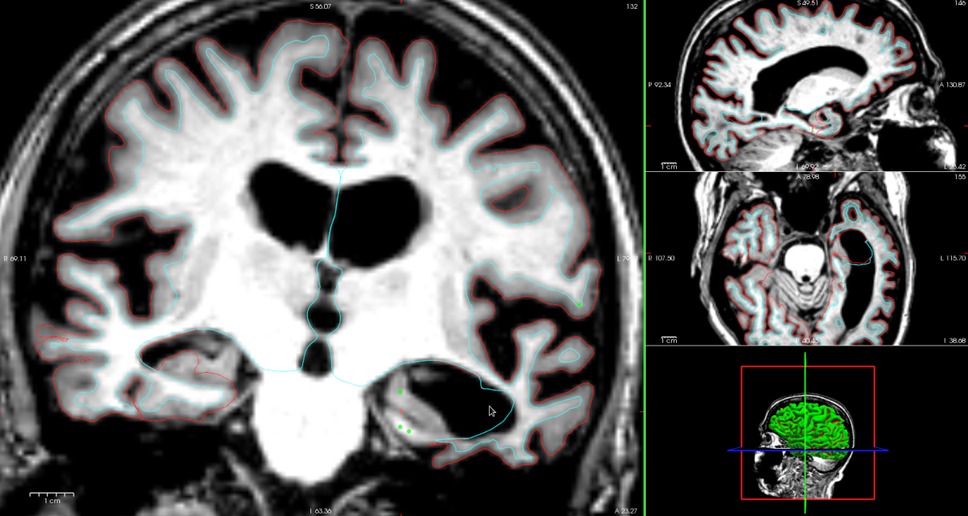
I have a set of subjects with moderate to severe temporal lobe atrophy. Surfaces are generally good everywhere except for portions of temporal lobes. It seems like control points are usually helpful in cases with problematic temporal lobes (ie. https://mail.nmr.mgh.harvard.edu/pipermail//freesurfer/2014-July/039596.html), but I'm hesitant to add too many control points in areas with limited/no wm (aka severely atrophied areas) for risk of misidentifying gm as wm. My goal is to run a cortical thickness analysis and look specifically at the temporal lobes, so I want to process/troubleshoot the subjects in as similar a manner as possible and produce generally accurate thickness maps.
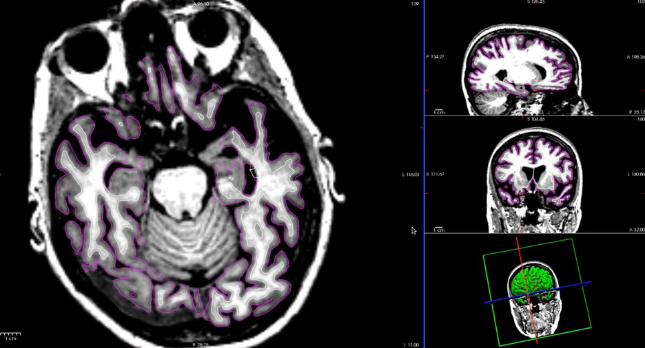
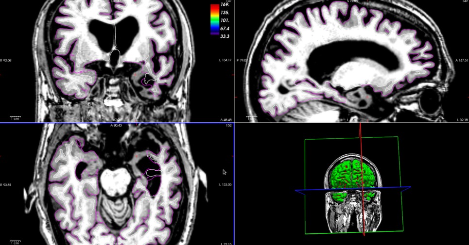
Attached are three screenshots: 1. tl_subj_A.jpeg : a subject with cp added across several slices and -autorecon2-cp -autorecon3 reprocessing; pial missing some gm in both lobes 2. tl_subj_B.jpeg : a subject after standard recon-all; generally good wm/gm recognition; good wm recognition but poor gm recognition in temporal lobes 3. tl_subj_C.jpeg : a subject after standard recon-all; generally similar to tl_subj_B.jpeg, but surfaces really went rogue in part of the atrophied lobe
Questions: - Is there a combination of flags that would help identify gm in the temporal lobes of most subjects while maintaining wm accuracy? - Any suggestions for adding cp (or other edits) in a semi-standard/efficient way that would still allow for a valid cortical thickness analysis? - What happened to the surfaces in subjects like the one in tl_subj_C.jpeg? - In terms of checking output quality for a cortical thickness analysis, would it be most useful to use QA Tools (https://surfer.nmr.mgh.harvard.edu/fswiki/QATools) or another method such as creating histograms from one of the stats files and looking for outliers? - (In general) How much quality checking / editing is appropriate for a cortical thickness study?
Thank you for any suggestions and/or resources!!
Sara