
13 Jun
2014
13 Jun
'14
8:57 a.m.
Dear all,
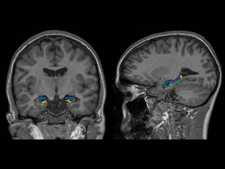
I'm running the GEMS tool in Freesurfer 5.3 to measure the volumes of hippocampal subfields in subjects between 9 and 14 years of age with 3T T1-weighted MRI scans.
I noticed that a blackline is consistently drawn around the surface of the subfields (see attached figure) and was wondering whether this is normal.
Since the tool has not been validated in young subjects I would like to know if the black line observed in the figure shows that the segmentation has not been carried out correctly.
Any help will be greatly appreciated.
Many thanks,
Joana