
28 Aug
2020
28 Aug
'20
7:27 p.m.
Hello,
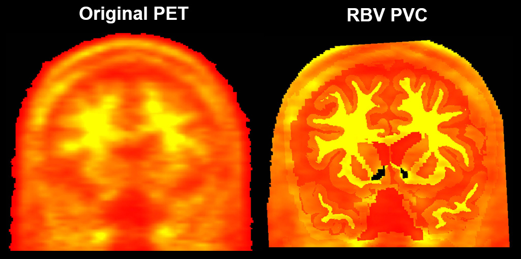
I’ve been running RBV partial volume correction on some amyloid PET data recently using mri_gtmpvc, and for ~90% of subjects the resulting rbv.nii.gz looks as expected with the exception of negative values in the bilateral accumbens (see attached image, thresholded from 0-4000 in scanner units). Any ideas why this might be happening?
This is the command I’ve been using: mri_gtmpvc --i $PETimg \ --reg $identity_mtrx \ --seg $FSdir/${subject}/mri/gtmseg.mgz \ --psf 6 --default-seg-merge --auto-mask PSF .01 --rbv \ --no-rescale --threads 8 \ --o $outname
Thanks very much,
Dan