
16 Jul
2008
16 Jul
'08
3:43 p.m.
Hello Surfers,
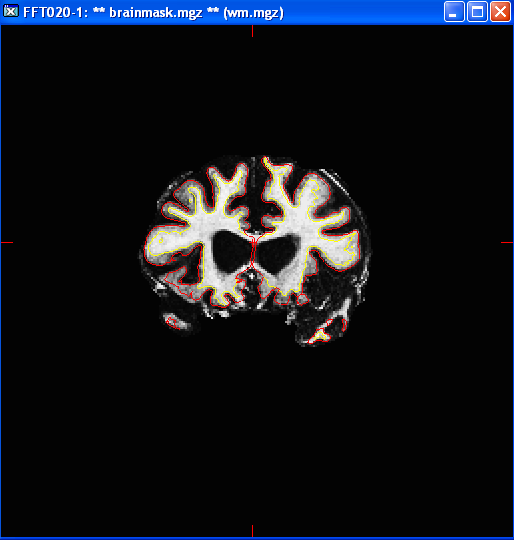
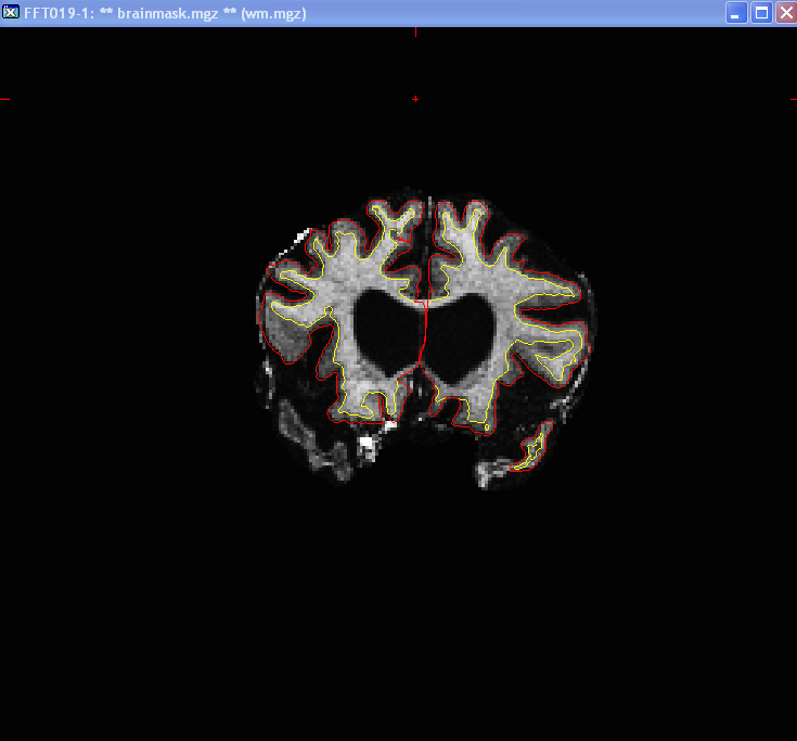
I am trying to FreeSurfer process a group of subjects from our Frontal Temporal Lobar Dementia project that were scanned on our 4T Siemens MRI. So far this endeavor has only created long hours of edits due to the absence of any visible White Matter in the Frontal and/or Temporal Poles. I have included a couple pictures of FS surface estimate on two of our many problematic subjects. I am wondering if you have any solutions or input to help FS include these regions of great atrophy.
Thanks in advance for any input on this tedious issue.
Sky R.