
Hi,
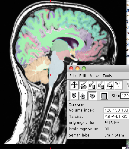
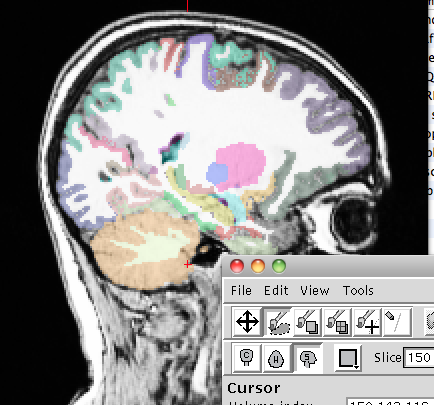
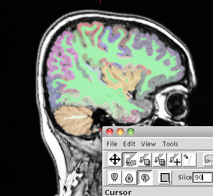
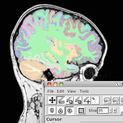
We have a dataset of 80 subjects whose T1 MPR images (1x1x1 resolution, acquired on a Siemens 1.5T scanner) I processed with recon-all, and the segmentation of cerebellum looks questionable in nearly all of them. In some cases it is clear that non-cerebellar tissue is being labeled as cerebellum, and in other cases it is clear that the cerebellum is not being fully labeled (in both types of scenarios I confirmed by checking the voxels in all orientations).
Attached are some example snapshots. In one, you can see that part of the brainstem is being labeled as cerebellum; in another, the cerebellum has not been fully labeled; and in the other two, there are disconnected "islands" labeled as cerebellum that should not be. (The suboptimal quality of the images in general is due to difficulties with scanning our young and abnormal subjects.)
Can anybody suggest any fixes other than manual editing? I can upload a subject if requested.
Thank you,
Warren