
Dear all,
I would like to show some very frequent and typical cases of segmentation problems in my bunch of data of normal subjects (after recon-all), of which I couldn't find a description in the wiki ( http://surfer.nmr.mgh.harvard.edu/fswiki/FreeviewGuide/FreeviewWorkingWithDa... ).
I am therefore not sure if these types of error are serious and will affect later results like thickness, curvature, GWR, etc.
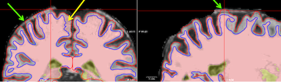
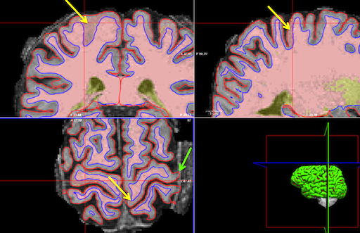
Here are the examples (c.f. attached screenshots from Freeview)
1) In the primary sensory-motor cortex it frequently happens that the "?h.white" line lies several mm within the wm border (c.f. yellow arrow), I did not observe this behavior in other places.
Would I need to edit that?
2) Sometimes the "?h.pial" line includes also dura or parts of the skull (green arrow).
Is it necessary to edit that as well?
3) Another question concerns the yellow parts of the wm which sometimes are much larger than the ventricles and also include both caudate nuclei (not shown).
Is this a problem?
Thanks in advance!!
Markus