
Thank you so much, Anastasia! Your comments help a lot and save a huge amount of time.. Indeed, I'm afraid, I have more questions.
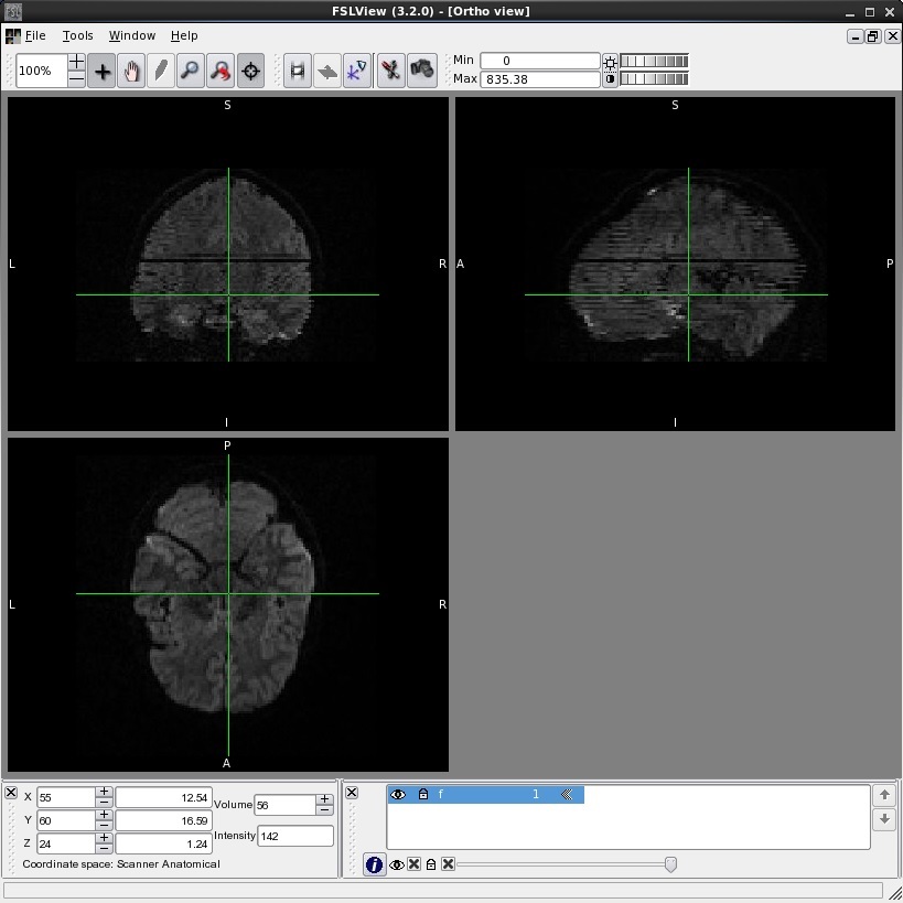
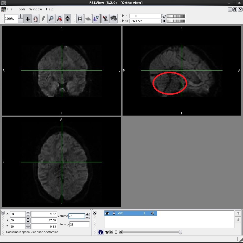
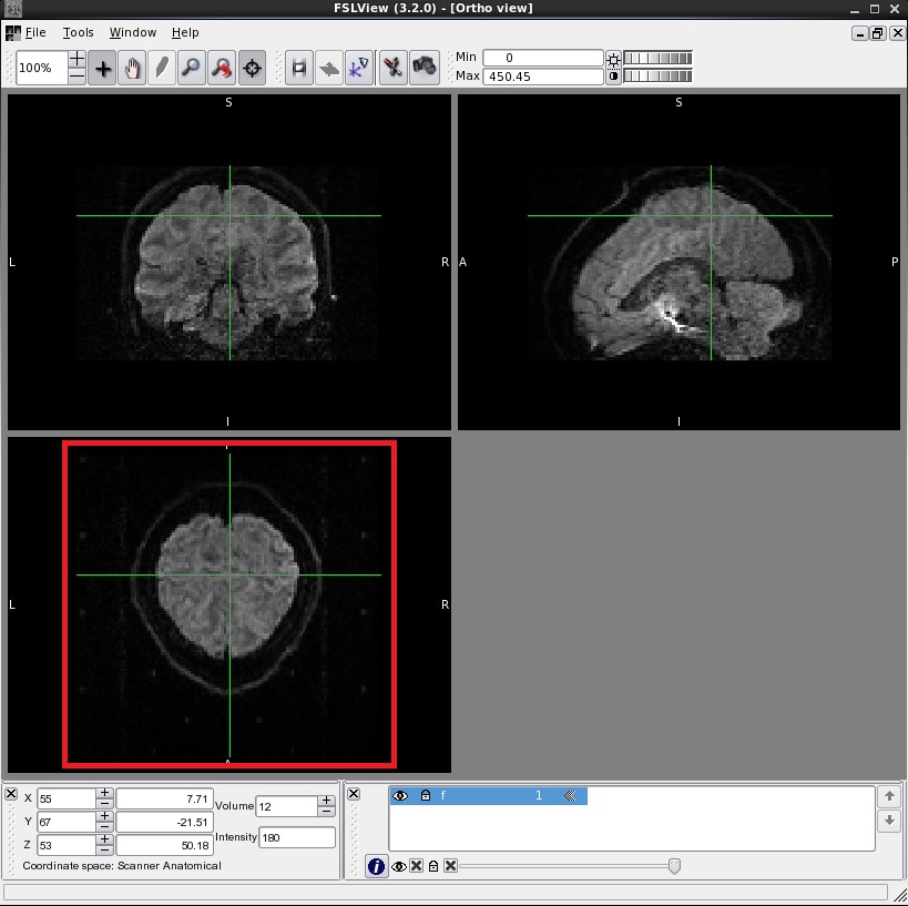
Until now I have only done what you proposed in 3. I observed 5 types of problems in a total of 12 (out of 78) time points: The first is what you described: A dark line referring to one slice that is much darker than its neighbors. I was happy to find this in only 1-2 volumes of all. The second is the same but the slices being only slightly darker than their neighbors. I guess it's just up to me what to do here, just need to make a decision. This happens more often. The third is a distortion of a sequence of slices. It cannot be a movement artifact (I think) because it is going back and forth with every slice. In attached picture "1" you can clearly detect the type 1 problem (the dark line) but also this third problem over almost the entire volume. It is as if the slices were not properly aligned to each other there. This is the most abundant problem type. The fourth type of problem is similar to the first 3, only that the lines don't go through the entire brain, which seems weird..I only detected this once, see picture "2". The last one is a background artifact that is visible in the z-axes, see picture "3", I'm afraid it will mess up this scan completely..I also detected this only once.
As I said, I was pretty happy in the beginning that I only saw these 1-2 volumes that were very dark. However, I also had a look at the dmri/dwi files after the eddy and simple motion correction step and unfortunately all of the mentioned problems persist. :( Especially concerning the third type of problem this is really a pain.
So it seems, I have to remove a lot of volumes and/or need to take some of the subjects completely out of our analysis. For this I would like to know: / //When I remove 1,2..10, 20 volumes, how does this impact the calculations of the tensor or the ball-and-stick parameters? I thought to find some repetitions in the bvec-files but it seems there are really 60 unique directions in our dwi-scans and I wonder why I can just remove one (or more/how many per subject?)./// / //Were problem types 3-5 reported by someone else before and can I do something about them apart from removing the volumes?/
On an additional note: I told you much earlier that I had to reinitialize some tracts of some subjects as they were not at all or badly reconstructed. However, this does not seem to correlate with these problems here so I wonder if TRACULA maybe is not so much effected by these problems after all?
Thank you! Vincent
Am 10/14/2013 5:12 PM, schrieb Anastasia Yendiki:
Hi Vincent,
- It's a pain, isn't it? I sympathize but looking at the data is always a
good idea. Since you'll see all 3 views in fslview, the dark slices will look like dark lines in two of these views, so you don't have to scroll through slices. There's a couple of things you can do about motion, for example you can remove the DWI volumes that have the artifacts. But you'll have to make sure you're not doing this more for one group than the other.
- What I usually look at is dlabel/diff/aparc+aseg.bbr.nii.gz, overlaid
on dmri/dtifit_FA.nii.gz. And yes, I do this for all subjects and yes, it takes a while. Listening to music helps.
Let me know if you have more questions! a.y
On Mon, 14 Oct 2013, Vincent Brunsch wrote:
Hi Anastasia!
I will go backwards with increasing difficulty to understand everything:
I see, yes this would not make sense. Thank you for the explanation!
With fslview I will have to run through 393,120 slices, then. (72 slices
in z-direction for our 70 DWI scans for tp1 and tp2 for each of our 39 subjects) I will go dwi image by dwi image running through the 72 slices rather fast. Just to make sure I am not wasting a lot of time: is this what I should do? If I detect slices that are much darker than their neighbors, will I need to exclude this subject for the analysis or can I do something about it?
- Yes, I use bbregister and I also use the anatomical T1 weighted scan to
extract the brain mask (usemaskanat=1). To make sure that everything is alright, I would go slice by slice in tkmedit with tkmedit [subject] brainmask.mgz -surfs -aparc+aseg where [subject] would be the base AND both longitudinal runs. I understand that I need to check if white matter is where it should be, if the cortical and pial surfaces are where they should be and if the labelling is correct. Again, as this will take probably even longer than 3. I would like to make sure this is the right thing to do before starting the quality check.
Thanks again! Vincent
Am 10/11/2013 2:13 PM, schrieb Anastasia Yendiki:
Hi Vincent - I'll take on the tracula-related parts:
- For tracula, the part of the recon-all output that matters is the
aparc+aseg. The surfaces will play a role only the DWI-to-T1 registration (assuming you opt to use bbregister).
- It's important to check your DWI data for obvious motion artifacts,
(slices that are much darker than their neighbors). Right now this has to be done visually, but it's on my list to produce some motion metrics as part of the preprocessing.
- The ball-and-stick model (that bedpostx fits to your data) is used by
the tractography algorithm in tracula, but there are no stats produced on the parameters of that model currently. That's something that can be added in the future as well. Note though that it wouldn't make sense to just average f1 or f2 over the pathway, because compartment 1 in one voxel may correspond to compartment 2 in some other voxel.
Hope this helps, a.y
On Fri, 11 Oct 2013, vbrunsch@nmr.mgh.harvard.edu wrote:
Dear Freesurfer experts,
I want to do a quality check on our imaging data. I used the longitudinal stream for SBA and as the first step for the longitudinal white matter analysis with TRACULA. We had two time points in our study and thus, in the freesurfer output directory there are 5 folders per subject (2 cross-sectional runs, 2 long runs and the base).
- Would you recommend to use (all of) the QA_TOOLS on all of these 5
folders per subject for the SBA? 2. Independent of the previous question, for the longitudinal version of TRACULA would you recommend to use (all of) the QA_TOOLS on the freesurfer base folder only / additional folders? 3. In addition to the late visual check for well reconstructed pathways with freeview, is there another automated possibility to check the quality of the diffusion weighted images beforehand/do you think this is necessary?
- On another note: If I understand correctly, in TRACULA bedpostX is used
to reconstruct the pathways but then the mean over the voxels that were hit (by the MCMC sampling of the paths) of measures from the tensor model are taken as outputs. I wonder, is there also the possibility of using the partial volumes f1, f2,.. as output measures?
Best, Vincent _______________________________________________ Freesurfer mailing list Freesurfer@nmr.mgh.harvard.edu https://mail.nmr.mgh.harvard.edu/mailman/listinfo/freesurfer
Freesurfer mailing list Freesurfer@nmr.mgh.harvard.edu https://mail.nmr.mgh.harvard.edu/mailman/listinfo/freesurfer