
hi Freesurfer team,
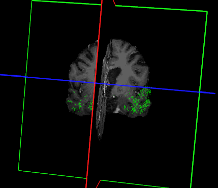
I tried to use less number of control points and tried to keep them in wm as much as I could guess the white matter boundary. Then I ran autorecon2-cp and autorecon3. But the white matter surfaces didn't expand at all. [cid:41bb92ba-7624-48f8-b1da-7486e1f055aa] Anything I am missing?
Regards, Manorama ________________________________ From: freesurfer-bounces@nmr.mgh.harvard.edu freesurfer-bounces@nmr.mgh.harvard.edu on behalf of Kadwani, Manorama MKADWANI@mgh.harvard.edu Sent: Thursday, May 27, 2021 5:56 PM To: Freesurfer support list freesurfer@nmr.mgh.harvard.edu Subject: Re: [Freesurfer] Using control points for intensity normalization
I see, it does look like they are in the cortex in 3D view. Although I tried to keep them in white matter in 2D planes as possible. I will try deleting some. Thank you, Manorama ________________________________ From: freesurfer-bounces@nmr.mgh.harvard.edu freesurfer-bounces@nmr.mgh.harvard.edu on behalf of Douglas N. Greve dgreve@mgh.harvard.edu Sent: Thursday, May 27, 2021 5:41 PM To: freesurfer@nmr.mgh.harvard.edu freesurfer@nmr.mgh.harvard.edu Subject: Re: [Freesurfer] Using control points for intensity normalization
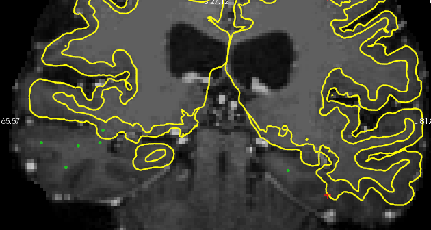
Generally, control points are the way to go here. It looks like you have put a bunch of them in cortex though
On 5/21/2021 12:14 PM, Kadwani, Manorama wrote: Hi,
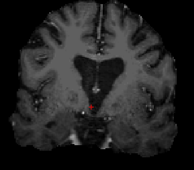
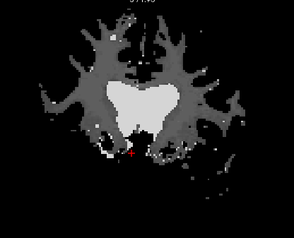
I am trying to correct the white matter segmentation in the second image shown below. It doesn't include chunks of the temporal lobe with low intensity on both hemispheres. [cid:part1.CCDF94B1.7201CC50@mgh.harvard.edu][cid:part2.FC9BA22F.8EE5EBBF@mgh.harvard.edu] I placed many control points on every slice in the brainmask.mgz file. Then I re-ran recon-all using -autorecon2-cp argument. But the white matter volume did not grow into those regions with control points. The process completed without any errors. [cid:part3.8EBAACCD.C13C0D60@mgh.harvard.edu] Any idea on how I can include those regions in white matter?
I also tried to use 'mri_segment' for lowering the white matter low limit to 60. That did include some parts of the missing lobes, but also included grey matter from frontal and parietal lobes. So, that approach was not ideal.
Thank you, Manorama
_______________________________________________ Freesurfer mailing list Freesurfer@nmr.mgh.harvard.edumailto:Freesurfer@nmr.mgh.harvard.edu https://mail.nmr.mgh.harvard.edu/mailman/listinfo/freesurfer