
Hi Bruce,
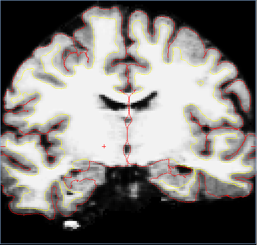
I was thinking it could be quality as well, since I asked about that in a previous question where my group compared 3T and 1.5T data from the same subject. Unfortunately we only obtained 1 scan for all of our subjects and nearly all of our data is from a 1.5T scanner. I've attached coronal, axial, and sagittal views with aseg+aparc, and one image without aseg+aparc for a WM segmentation check. Thanks for your help!
Jessica
On Thu, Jul 28, 2011 at 11:39 AM, Bruce Fischl fischl@nmr.mgh.harvard.eduwrote:
Hi Jessica
the problem may be data quality! At 1.5T with a volume coil we would typically get 2 scans to average. A TR of 14 is pretty short and will reduce the SNR, and a TE of 6 is quite long and will reduce contrast. Can you send us a tif of one slice to look at?
Bruce
On Thu, 28 Jul 2011, Jessica Liu wrote:
Hi Martin,
When I did longitudinal analyses in the past, the output was very strange even though I think I did everything correctly. If it's not too much trouble for you, could you please take a look at my data and tell me what I've done wrong? Since the files are so large, how would I send over my data? Also, I'm using is a head coil 1.5T single-channel GE scanner. The sequence I'm using is 3D SPGR with TR = 14 msec, TE = 6.2 msec, TI = 450.0, FOV 24 cm, data acquisition matrix = 256x192, and 124 slices with 1.6 mm thickness. Your help is truly appreciated!
Jessica
On Thu, Jul 28, 2011 at 7:37 AM, Martin Reuter < mreuter@nmr.mgh.harvard.edu> wrote: Hi Jessica,
 I'd recommend to run the -base and the -long on these and see what
 happens. If you then still see these differences, maybe we can take a
 look at your data to figure out what causes this. It could be that
the WM segmentation fails somewhere. Anyway, the longitudinal stream should help fix these things.
 Are these mprages or multi echo mprages? 3T? what coil?

 Best, Martin
On Wed, 2011-07-27 at 18:01 -0700, Jessica Liu wrote:
Hi Bruce,
Well for one of the subjects, the total temporal lobe volume from the first scan was 149937 mm3, and the second scan's total temporal lobe volume was 183121 mm3. These numbers were found by summing the volumes from the entorhinal, fusiform, parahippocampal, temporal pole, transverse temporal, and the inferior, middle, and superior temporal sections found in the lh.aparc.stats, rh.aparc.stats, and wmparc.stats for the respective scans. Hippocampus volumes were found by summing the right and left parts found in aseg.stats.
Jessica
On Wed, Jul 27, 2011 at 5:37 PM, Bruce Fischl fischl@nmr.mgh.harvard.edu wrote: how are you computing the 20% difference? We definitely don't see this, and Martin has done extensive testing of repeatability. Bruce
On Wed, 27 Jul 2011, Jessica Liu wrote: Hi Martin, Thanks for getting back to me. Actually I have two young controls who have 20% differences between two scans a day apart. Visually, there are not abnormalities for the surfaces and hippocampus in both subjects i.e. on tkmedit, the color of the hippocampus area (yellow) looks alright to me. I don't see any motion artifacts either. Jessica On Wed, Jul 27, 2011 at 12:15 PM, Martin Reuter <mreuter@nmr.mgh.harvard.edu> wrote: Hi Jessica, I don't think this is normal. Is this a single subject? Of course in a single subject lots of stuff can go wrong. Are the surfaces correct? And the hippo label? Are there motion artifacts in the image etc. Anyway, you should process this with the longitudinal stream: http://freesurfer.net/fswiki/**LongitudinalProcessing<http://freesurfer.net/fswiki/LongitudinalProcessing> which should increase repeatability. Best, Martin On Wed, 2011-07-27 at 09:01 -0700, Jessica Liu wrote: > Hi, > > We found 20% differences in temporal lobe brain volume and ca. 5% > difference in the hippocampus volume between two data sets of a young > normal volunteer scanned 2 days apart. > > We use the one-step 19 hours recon-all -all procedure and directly sum > select values taken from the lh.aparc.stats, rh.aparc.stats, and > wmparc.stats. The values summed were based on information given from > https://mail.nmr.mgh.harvard.**edu/pipermail/freesurfer/2007-**April/005000.htmlhttps://mail.nmr.mgh.harvard.edu/pipermail/freesurfer/2007-April/005000.html
            >
            > My question is, are these observations normal for
            Freesurfer?  Any
            > comments are greatly appreciated.  Thanks!
            > --
            > Pom & Jessica
            >
            >
            >
            > ______________________________**_________________
            > Freesurfer mailing list
            > Freesurfer@nmr.mgh.harvard.edu
            >
            https://mail.nmr.mgh.harvard.**edu/mailman/listinfo/**
freesurfer https://mail.nmr.mgh.harvard.edu/mailman/listinfo/freesurfer
            The information in this e-mail is intended only for
            the person to whom it is
            addressed. If you believe this e-mail was sent to you
            in error and the e-mail
            contains patient information, please contact the
            Partners Compliance HelpLine at
            http://www.partners.org/**complianceline<http://www.partners.org/complianceline>. If the e-mail
            was sent to you in error
            but does not contain patient information, please
            contact the sender and properly
            dispose of the e-mail.