
External Email - Use Caution
Dear Freesurfer experts, I have two groups of subjects (healthy and patients). The patients have different degrees of atrophy. Looking into the individual scans, the subjects are largely different in the space between the sulcuses and the size of ventricles. My questions are: 1. Is there any way to correct for these differences in the surface based analysis? 2. Including eTIV covariate does it help in this case? 3. Does the resampling to "fsaverage" overcome the challenge of these differences between the subjects?
I appreciate any advice, John

Hi John
why do you want to correct for them? They likely represent a pathological process, so correcting for them may remove whatever effects you are looking for, no? The space between the sulci and the ventricle volume won't directly affect surface-based analysis, but of course the sulcal widening likely reflects cortical atrophy that will be
cheers Bruce
On Wed, 11 Jul 2018, John Anderson wrote:
External Email - Use Caution
Dear Freesurfer experts, I have two groups of subjects (healthy and patients). The patients have different degrees of atrophy. Looking into the individual scans, the subjects are largely different in the space between the sulcuses and the size of ventricles. My questions are:
- Is there any way to correct for these differences in the surface based analysis?
- Including eTIV covariate does it help in this case?
- Does the resampling to "fsaverage" overcome the challenge of these differences between the
subjects?
I appreciate any advice, John
External Email - Use Caution
Dear Dr Bruce,
Thank you for the response. Indeed, I was completely wrong, these represent a pathology and should not correct for!
Kindly, I have one additional question not relevant to the subject of this email. Is there any way in Freesurfer to create mask for the regions between the sulcuses. I want to compute PET signal in these regions and normalize for it.
Thank you so much! John
Hi John
why do you want to correct for them? They likely represent a pathological process, so correcting for them may remove whatever effects you are looking for, no? The space between the sulci and the ventricle volume won't directly affect surface-based analysis, but of course the sulcal widening likely reflects cortical atrophy that will be
cheers
Bruce
‐‐‐‐‐‐‐ Original Message ‐‐‐‐‐‐‐ On July 11, 2018 11:37 AM, John Anderson John.anderso@protonmail.com wrote:
Dear Freesurfer experts, I have two groups of subjects (healthy and patients). The patients have different degrees of atrophy. Looking into the individual scans, the subjects are largely different in the space between the sulcuses and the size of ventricles. My questions are:
- Is there any way to correct for these differences in the surface based analysis?
- Including eTIV covariate does it help in this case?
- Does the resampling to "fsaverage" overcome the challenge of these differences between the subjects?
I appreciate any advice, John
I suppose you could look for voxels that outside of the cortex and adjacent to it On Wed, 11 Jul 2018, John Anderson wrote:
External Email - Use Caution
Dear Dr Bruce,
Thank you for the response. Indeed, I was completely wrong, these represent a pathology and should not correct for!
Kindly, I have one additional question not relevant to the subject of this email. Is there any way in Freesurfer to create mask for the regions between the sulcuses. I want to compute PET signal in these regions and normalize for it.
Thank you so much! John
Hi John
why do you want to correct for them? They likely represent a pathological process, so correcting for them may remove whatever effects you are looking for, no? The space between the sulci and the ventricle volume won't directly affect surface-based analysis, but of course the sulcal widening likely reflects cortical atrophy that will be
cheers
Bruce
‐‐‐‐‐‐‐ Original Message ‐‐‐‐‐‐‐ On July 11, 2018 11:37 AM, John Anderson John.anderso@protonmail.com wrote:
Dear Freesurfer experts,I have two groups of subjects (healthy and patients). The patients have different degrees of atrophy. Looking into the individual scans, the subjects are largely different in the space between the sulcuses and the size of ventricles. My questions are:
- Is there any way to correct for these differences in the surface based analysis?
- Including eTIV covariate does it help in this case?
- Does the resampling to "fsaverage" overcome the challenge of these differences between the
subjects?
I appreciate any advice, John
External Email - Use Caution
Thank you so much Dr Bruce,
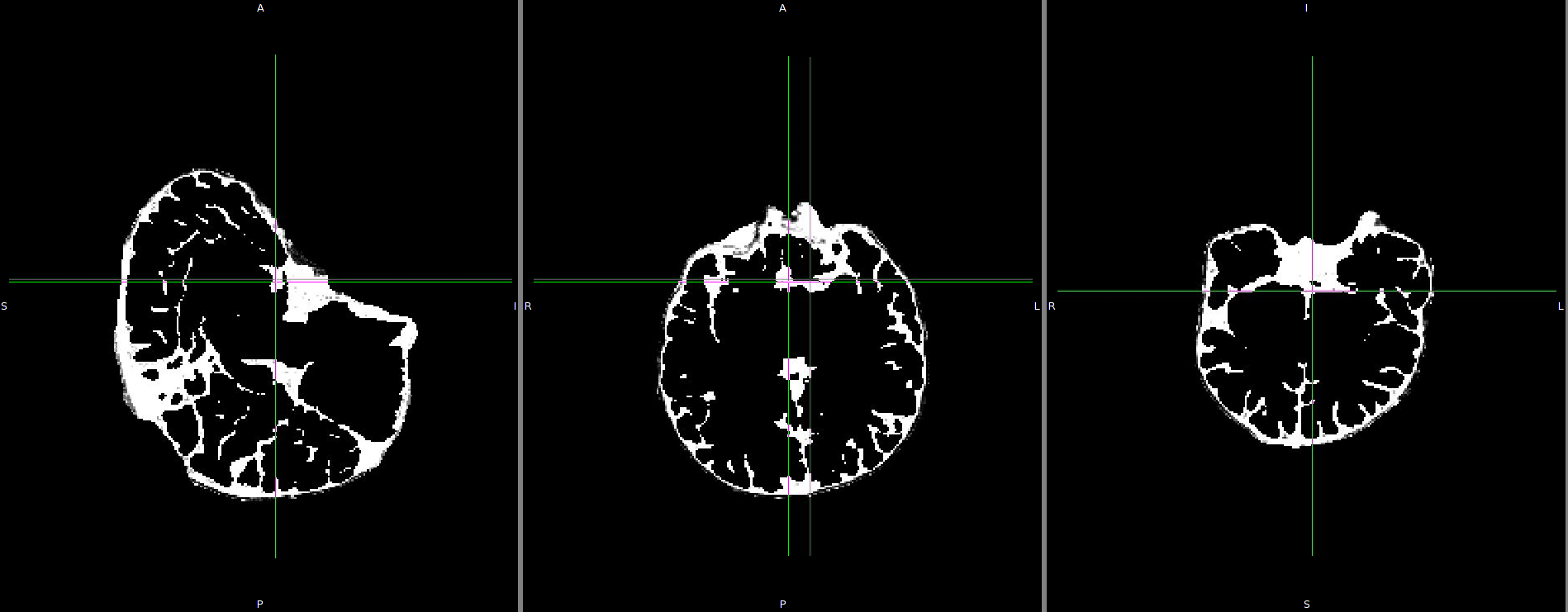
I tried to create this maks in may different ways. Finally, and following many attempts . I tried these commands: mri_binarize --i aparc.DKTatlas+aseg.nii.gz --min 0 --max .1 --o reversed_mask.nii --binval 1 mri_convert $SUBJECTS_DIR/$Subj/mri/brainmask.mgz brainmask.nii mri_mask brainmask.nii reversed_mask.nii sulcus.nii.gz
Attached is a snapshot for the output. Please are these steps correct? I would appreciate any suggestion that would improve this mask.
John
I suppose you could look for voxels that outside of the cortex and adjacent to it On Wed, 11 Jul 2018, John Anderson wrote:
‐‐‐‐‐‐‐ Original Message ‐‐‐‐‐‐‐ On July 11, 2018 2:03 PM, John Anderson John.anderso@protonmail.com wrote:
Dear Dr Bruce,
Thank you for the response. Indeed, I was completely wrong, these represent a pathology and should not correct for!
Kindly, I have one additional question not relevant to the subject of this email. Is there any way in Freesurfer to create mask for the regions between the sulcuses. I want to compute PET signal in these regions and normalize for it.
Thank you so much! John
Hi John
why do you want to correct for them? They likely represent a pathological process, so correcting for them may remove whatever effects you are looking for, no? The space between the sulci and the ventricle volume won't directly affect surface-based analysis, but of course the sulcal widening likely reflects cortical atrophy that will be
cheers
Bruce
‐‐‐‐‐‐‐ Original Message ‐‐‐‐‐‐‐ On July 11, 2018 11:37 AM, John Anderson John.anderso@protonmail.com wrote:
Dear Freesurfer experts, I have two groups of subjects (healthy and patients). The patients have different degrees of atrophy. Looking into the individual scans, the subjects are largely different in the space between the sulcuses and the size of ventricles. My questions are:
- Is there any way to correct for these differences in the surface based analysis?
- Including eTIV covariate does it help in this case?
- Does the resampling to "fsaverage" overcome the challenge of these differences between the subjects?
I appreciate any advice, John

On 7/11/2018 11:37 AM, John Anderson wrote:
External Email - Use Caution
Dear Freesurfer experts, I have two groups of subjects (healthy and patients). The patients have different degrees of atrophy. Looking into the individual scans, the subjects are largely different in the space between the sulcuses and the size of ventricles. My questions are: 1. Is there any way to correct for these differences in the surface based analysis? What kind of correction? I'm not sure I understand what the problem is. 2. Including eTIV covariate does it help in this case? No, since the skull does not change with atrophy 3. Does the resampling to "fsaverage" overcome the challenge of these differences between the subjects? See #1
I appreciate any advice, John
_______________________________________________ Freesurfer mailing list Freesurfer@nmr.mgh.harvard.edumailto:Freesurfer@nmr.mgh.harvard.edu https://mail.nmr.mgh.harvard.edu/mailman/listinfo/freesurfer
freesurfer@nmr.mgh.harvard.edu
-
 Bruce Fischl
 -
 Greve, Douglas N.,Ph.D.
 -
 John Anderson