
Hi All,
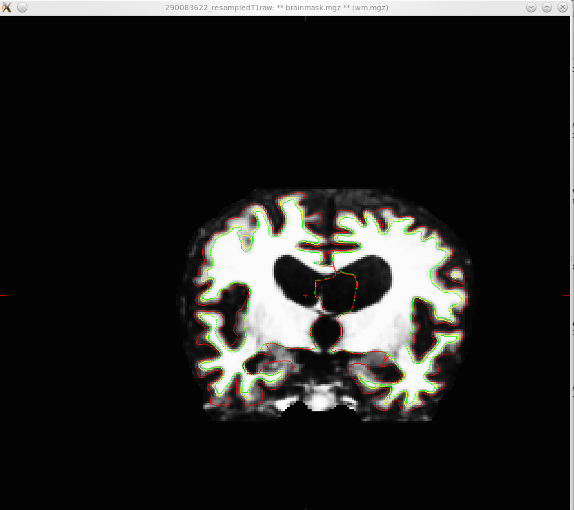
When use tkemedit to view my results of autorecon2, I got the attached image. I think this is not correct(I am not very sure). Is there any one know why this happens and any way to solve this?
Thanks a lot.
Best, Xiaoying

Hi Xiaoying,
do you mean the ventricles? They are pretty big and may not have been labeled accurately. The cortex is hard to tell the way you have it windowed.
cheers Bruuce On Fri, 10 Dec 2010, Xiaoying Tang wrote:
Hi All,
When use tkemedit to view my results of autorecon2, I got the attached image. I think this is not correct(I am not very sure). Is there any one know why this happens and any way to solve this?
Thanks a lot.
Best, Xiaoying
Yes--- the ventricles. I am guessing whether it is because the brain is of subject who has some kind of disease. My goal is to segment the subcortical structures like Basal Ganglia. Does this affect my segmentation follows?
Best, Xiaoying
2010/12/10 Bruce Fischl fischl@nmr.mgh.harvard.edu
Hi Xiaoying,
do you mean the ventricles? They are pretty big and may not have been labeled accurately. The cortex is hard to tell the way you have it windowed.
cheers Bruuce
On Fri, 10 Dec 2010, Xiaoying Tang wrote:
Hi All,
When use tkemedit to view my results of autorecon2, I got the attached image. I think this is not correct(I am not very sure). Is there any one know why this happens and any way to solve this?
Thanks a lot.
Best, Xiaoying
The information in this e-mail is intended only for the person to whom it is addressed. If you believe this e-mail was sent to you in error and the e-mail contains patient information, please contact the Partners Compliance HelpLine at http://www.partners.org/complianceline . If the e-mail was sent to you in error but does not contain patient information, please contact the sender and properly dispose of the e-mail.
freesurfer@nmr.mgh.harvard.edu
-
 Bruce Fischl
 -
 Xiaoying Tang