
Hi all,
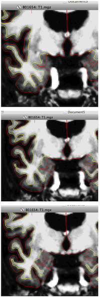
I have a question about what to me looks like an inaccurate surface reconstruction. I attach here 3 consecutive coronnal slices: in the first and second, the pial surface doesn't seem to follow the whole extent of the collateral sulcus on the right hemisphere (the location of the crosshair). Just eye balling it, it looks like there is a relatively deep sulcus there, but the pial surface doesn't go all the way in. In the third screen shot, 2mm posterior to the first, the sulcus is reconstructed the way I would expect it to.
Could you please give me some insights into the reason behind this? Is there a way of fixing it without touching the gray matter voxels? Please let me know if you need any additional information!
Thank you in advance, Sasa
freesurfer@nmr.mgh.harvard.edu
-
 Sasa Kivisaari