
Hi Bruce:
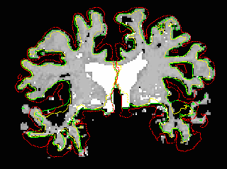
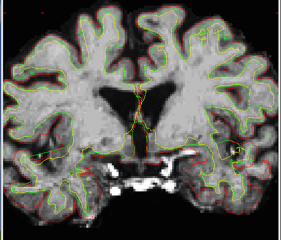
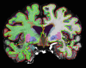
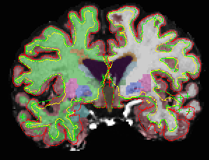
I looked at the brainmask.mgz and wm.mgz - obviously the pial is not included in superior & middle temporal regions in the brainmask.mgz and in the wm.mgz. The wm intensities are ranging from 96 - 106 in the missing superior & middle temporal regions in the brainmask.mgz. I have attached the brainmask.mgz, wm.mgz, aseg.mgz & aseg+aparc.mgz, all with pial, white and orig surfaces overlaid.
As mentioned earlier, I had control points added in wm of excluded temporal regions and ran this command again ( to fix pial surface ) :
recon-all -autorecon2-cp -autorecon3 -subjid subject
The final image did not include the excluded temporal region after opening up in:
tkmedit subject brainmask.mgz lh.white -aux T1.mgz -aux-surface rh.white
But gave me different values for entire regions in lh & rh aparc and aparc.a2005.stats - cov >1% in all regions. For example,
Before adding control point After adding control point COV
lh superior temporal(mm2) 3487 3102 8.3%
rh superior temporal (mm2) 3434 3370 4.9%
lh middle temporal(mm2) 3902 3001 18.5%
rh middle temporal (mm2) 3046 3528 10.4%
Another question:
if you run this command > recon-all -autorecon2-cp -autorecon3 -subjid subject < Do we run the entire subcortical segmentation as well?
I appreciate your suggestions.
Deepa.
Deepa Preeti Ramasamy, MD Research Assistant Buffalo Neuroimaging Center The Jacobs Neurological Institute Department of Neurology Buffalo General Hospital 100 High St. Buffalo, NY 14203 Work: 716-859-7038 E-Mail: dramasamy@bnac.net

Hi Deepa,
if you see the orig surface not include some of the wm that is correctly segmented in the wm.mgz (as in your first image) it means that the topology correction incorrectly removed some stuff. You'll need to manually correct the defect, which should be visible on the ?h.inflated.nofix surface.
cheers, Bruce
On Thu, 22 Mar 2007, deepa preeti wrote:
Hi Bruce:
I looked at the brainmask.mgz and wm.mgz - obviously the pial is not included in superior & middle temporal regions in the brainmask.mgz and in the wm.mgz. The wm intensities are ranging from 96 - 106 in the missing superior & middle temporal regions in the brainmask.mgz. I have attached the brainmask.mgz, wm.mgz, aseg.mgz & aseg+aparc.mgz, all with pial, white and orig surfaces overlaid.
As mentioned earlier, I had control points added in wm of excluded temporal regions and ran this command again ( to fix pial surface ) :
recon-all -autorecon2-cp -autorecon3 -subjid subject
The final image did not include the excluded temporal region after opening up in:
tkmedit subject brainmask.mgz lh.white -aux T1.mgz -aux-surface rh.white
But gave me different values for entire regions in lh & rh aparc and aparc.a2005.stats - cov >1% in all regions. For example,
Before adding controlpoint After adding control point COV
lh superior temporal(mm2) 3487 3102 8.3%
rh superior temporal (mm2) 3434 3370 4.9%
lh middle temporal(mm2) 3902 3001 18.5%
rh middle temporal (mm2) 3046 3528 10.4%
Another question:
if you run this command > recon-all -autorecon2-cp -autorecon3 -subjid subject < Do we run the entire subcortical segmentation as well?
I appreciate your suggestions.
Deepa.
Deepa Preeti Ramasamy, MD Research Assistant Buffalo Neuroimaging Center The Jacobs Neurological Institute Department of Neurology Buffalo General Hospital 100 High St. Buffalo, NY 14203 Work: 716-859-7038 E-Mail: dramasamy@bnac.net
freesurfer@nmr.mgh.harvard.edu
-
 Bruce Fischl
 -
 deepa preeti