
Hi Bruce:
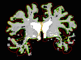
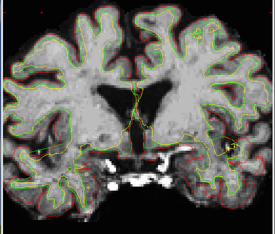
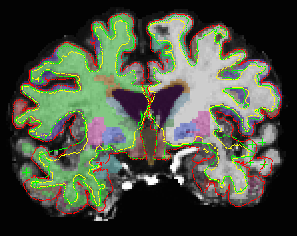
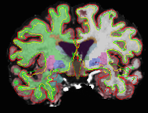
I looked at the brainmask.mgz and wm.mgz - obviously the pial is not included in superior & middle temporal regions in the brainmask.mgz and in the wm.mgz. The wm intensities are ranging from 96 - 106 in the missing superior & middle temporal regions in the brainmask.mgz. I have attached the brainmask.mgz, wm.mgz, aseg.mgz & aseg+aparc.mgz, all with pial, white and orig surfaces overlaid.
As mentioned earlier, I had control points added in wm of excluded temporal regions and ran this command again ( to fix pial surface ) :
recon-all -autorecon2-cp -autorecon3 -subjid subject
The final image did not include the excluded temporal region after opening up in:
tkmedit subject brainmask.mgz lh.white -aux T1.mgz -aux-surface rh.white
But gave me different values for entire regions in lh & rh aparc and aparc.a2005.stats - cov >1% in all regions. For example,
Before adding control point After adding control point COV
lh superior temporal(mm2) 3487 3102 8.3%
rh superior temporal (mm2) 3434 3370 4.9%
lh middle temporal(mm2) 3902 3001 18.5%
rh middle temporal (mm2) 3046 3528 10.4%
Another question:
if you run this command > recon-all -autorecon2-cp -autorecon3 -subjid subject < Do we run the entire subcortical segmentation as well?
I appreciate your suggestions.
Deepa.
Deepa Preeti Ramasamy, MD Research Assistant Buffalo Neuroimaging Center The Jacobs Neurological Institute Department of Neurology Buffalo General Hospital 100 High St. Buffalo, NY 14203 Work: 716-859-7038 E-Mail: dramasamy@bnac.net