
External Email - Use Caution
Hello,
I have run FreeSurfer v7.1.1 on the CBRAIN platform and I'm performing QC on the output using the information at the following page: https://secure-web.cisco.com/1oUroAUDs4KHQ2Dm_LBBIXd9QC0wayhlpqddWcLfLv3VRDC...
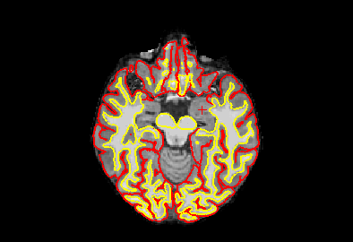
It seems though that for almost all of my output, a large portion of the grey matter is not being segmented (please see attached example).
I'm not sure if there is a solution to this or if the images will just need to be manually edited to fix this under segmentation. I would greatly appreciate any help or insight.
Thank you for your time and help,
Fatima

External Email - Use Caution
Hello,
The regions you have denoted with the cursor are not part of the cortex, and as such are not included in the pial/wm segmentation.
The 'un-segmented' areas are part of the medial wall. This is not a completly exhaustive list, but these structures are not classified as cortex and are not included in the pial/wm segmentation:
* Left-Accumbens-area * Left-Caudate * Left-Cerebral-White-Matter * Left-Lateral-Ventricle * Left-Pallidum Left-Putamen * Left-Thalamus * Left-VentralDC * Left-vessel * non-WM-hypointensities * Right-Accumbens-area * Right-Caudate Right-Cerebral-White-Matter * Right-Lateral-Ventricle * Right-Pallidum * Right-Putamen * Right-Thalamus * Right-VentralDC * Right-vessel * WM-hypointensities
Best, Jackson
________________________________ From: freesurfer-bounces@nmr.mgh.harvard.edu freesurfer-bounces@nmr.mgh.harvard.edu on behalf of Fatima Abboud fatme.abboud@mail.mcgill.ca Sent: Thursday, July 27, 2023 2:36 PM To: freesurfer@nmr.mgh.harvard.edu freesurfer@nmr.mgh.harvard.edu Subject: [Freesurfer] Grey Matter Under Segmentation
External Email - Use Caution
Hello,
I have run FreeSurfer v7.1.1 on the CBRAIN platform and I'm performing QC on the output using the information at the following page: MailScanner has detected a possible fraud attempt from "secure-web.cisco.com" claiming to be https://secure-web.cisco.com/1EHPx82EsEjkj9T33XQS4P1zIXXNGFz26wvoKD7rn-VizGl...https://secure-web.cisco.com/1oUroAUDs4KHQ2Dm_LBBIXd9QC0wayhlpqddWcLfLv3VRDC9_tqsqPsOHwN7q8fERPShD-l4u9jDhB36L-cYV2Z9nV5XN2nPDgE_8Ebs3zR-YzCp-rCSWTH_cTazVFS-zy4_Iy3VqNmS3H_Qp1R_5Eba1oPWvceO_8xUCkZzA9SVsDnQKESrw7O7YHYa41qmyHSueTb4g60PKhPOwJu7Fqrn3MXxaJyeG4latI9UpW-5M1HoeC4rekckgR7wpkEhLiqvE9LFu1cQd7g5kd98N3IFvF4L2ZkrE2EJOznzXlxAfVPxsVnxP4lKvBoMtRZ5GdO6w35FpTvrw197XemFWaQ/https%3A%2F%2Fsurfer.nmr.mgh.harvard.edu%2Ffswiki%2FFsTutorial%2FOutputData_freeviewV6.0
It seems though that for almost all of my output, a large portion of the grey matter is not being segmented (please see attached example).
I'm not sure if there is a solution to this or if the images will just need to be manually edited to fix this under segmentation. I would greatly appreciate any help or insight.
Thank you for your time and help,
Fatima
freesurfer@nmr.mgh.harvard.edu
-
 Fatima Abboud
 -
 Nolan, Jackson