
Dear Sir/Madam,
I am using freesurfer for 3T MRI analysis of visual cortical thickness in elderly patients with ophthalmic disease. We are having a bit of trouble with the accuracy of cortical segmentation in the visual cortex (namely the pericalcarine region) as the cortex is quite thin in our patients (compared to controls). You can see in the attached image that whilst there is smooth cortical segmentation in the peri-calcarine region, the cortex is not visible to the naked eye. If you have any ideas on improving accuracy, I would be very grateful.
Thanks Megha

Higher than 1mm resolution helps a lot. We use 0.7mm isotropic in the HCP.
Peace,
Matt.
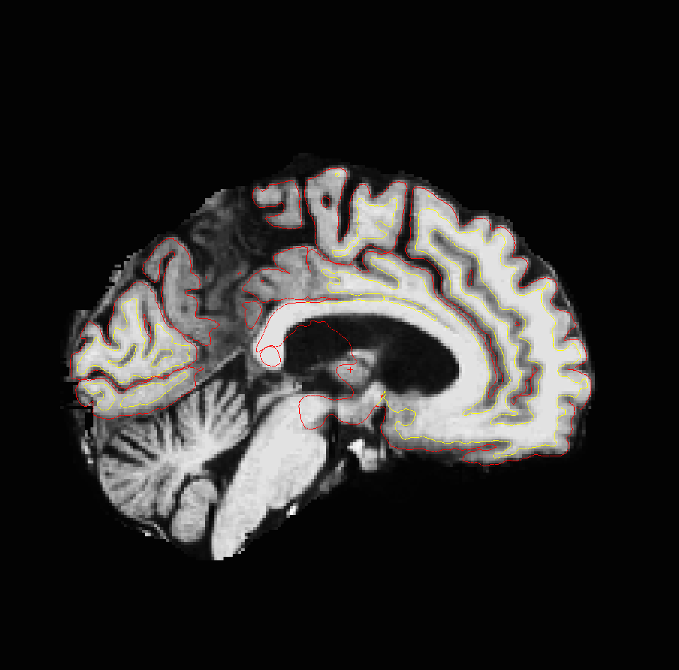
From: "Megha K." megha.ka@gmail.com Date: Monday, December 9, 2013 10:23 PM To: freesurfer@nmr.mgh.harvard.edu Subject: [Freesurfer] Occipital cortex segmentation
Dear Sir/Madam,
I am using freesurfer for 3T MRI analysis of visual cortical thickness in elderly patients with ophthalmic disease. We are having a bit of trouble with the accuracy of cortical segmentation in the visual cortex (namely the pericalcarine region) as the cortex is quite thin in our patients (compared to controls). You can see in the attached image that whilst there is smooth cortical segmentation in the peri-calcarine region, the cortex is not visible to the naked eye. If you have any ideas on improving accuracy, I would be very grateful.
Thanks Megha _______________________________________________ Freesurfer mailing list Freesurfer@nmr.mgh.harvard.edu https://mail.nmr.mgh.harvard.edu/mailman/listinfo/freesurfer The information in this e-mail is intended only for the person to whom it is addressed. If you believe this e-mail was sent to you in error and the e-mail contains patient information, please contact the Partners Compliance HelpLine at http://www.partners.org/complianceline . If the e-mail was sent to you in error but does not contain patient information, please contact the sender and properly dispose of the e-mail.

Hi Megha
looking at your images it's not clear to me where the gray/white boundary is. Do you think it should be further out? One thing you can try is (if the occipital white matter intensity in brain.mgz is <110) putting some control points in the interior of the white matter. Or as Matt suggests, higher resolution can help, but that may not be an option
cheers Bruce
On Tue, 10 Dec 2013, Megha K. wrote:
Dear Sir/Madam,
I am using freesurfer for 3T MRI analysis of visual cortical thickness in elderly patients with ophthalmic disease. We are having a bit of trouble with the accuracy of cortical segmentation in the visual cortex (namely the pericalcarine region) as the cortex is quite thin in our patients (compared to controls). You can see in the attached image that whilst there is smooth cortical segmentation in the peri-calcarine region, the cortex is not visible to the naked eye. If you have any ideas on improving accuracy, I would be very grateful.
Thanks Megha
freesurfer@nmr.mgh.harvard.edu
-
 Bruce Fischl
 -
 Matt Glasser
 -
 Megha K.