
9 Dec
2013
9 Dec
'13
11:23 p.m.
Dear Sir/Madam,
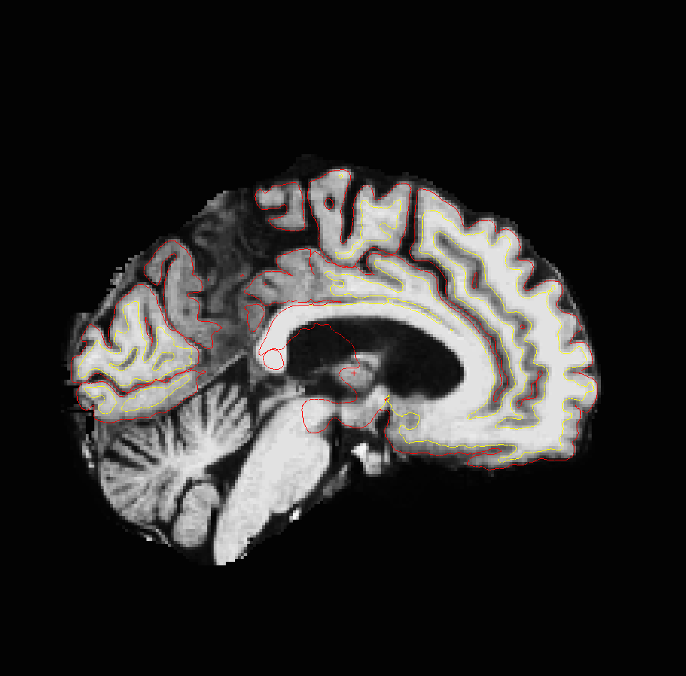
I am using freesurfer for 3T MRI analysis of visual cortical thickness in elderly patients with ophthalmic disease. We are having a bit of trouble with the accuracy of cortical segmentation in the visual cortex (namely the pericalcarine region) as the cortex is quite thin in our patients (compared to controls). You can see in the attached image that whilst there is smooth cortical segmentation in the peri-calcarine region, the cortex is not visible to the naked eye. If you have any ideas on improving accuracy, I would be very grateful.
Thanks Megha