
Hi Freesurfers,
I need some advice regarding the best approach to fixing some very large RECON errors in a highly lesioned brain.
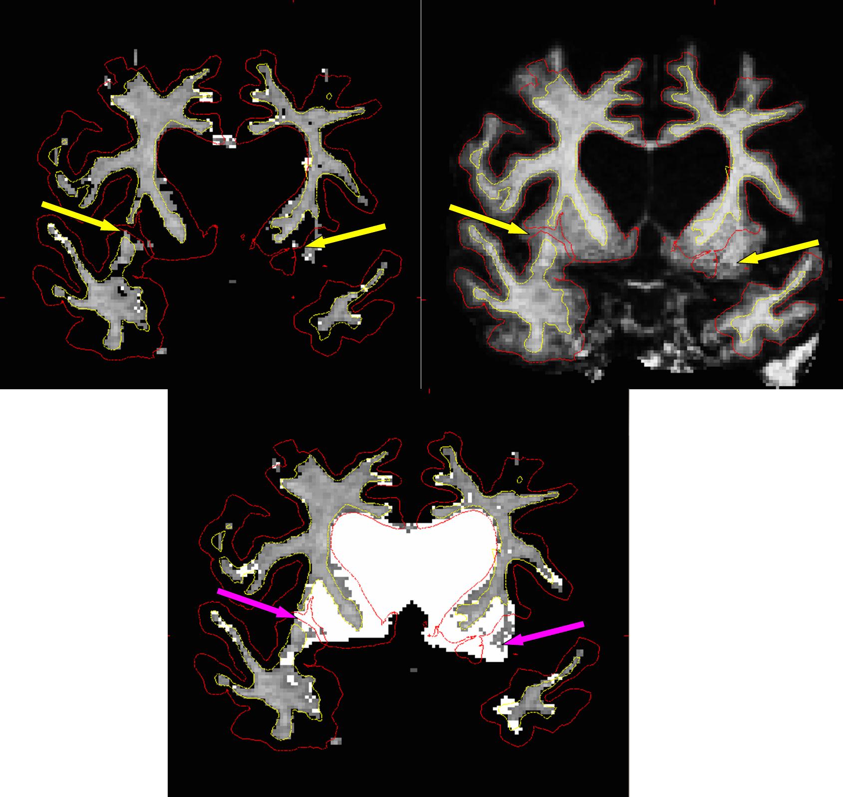
There are many problems in this RECON output, but I want to focus on one particularly bad one which is highlighted in the attached figure by yellow arrows but also have knock on effects in the ventricles. There are no examples in the tutorials or other docs that are quite like this. Can you tell me what the best approach to correcting this would be?
Are WM.MGZ edits, as highlighted by purple arrows, the right approach? I can't help but feel there is a lot of room for error with this approach and it could be extremely time-consuming due to the need to accurately trace the basal surface or the subcortical mass.
It is stated NOT to use control points in instances of white matter lesions. What about instances such as this? Clearly there is white and subcortical grey matter in this region - would placing control points in subcortical grey matter cause any problems? My take on control points is that it only informs the generation of the WM.MGZ image, which is not just white matter but generally all subcortical mass, and intensity changes instigated by them do not directly effect subcrotical parcellation - is this correct? If so, perhaps then control points would be OK to resolve this error.
Please advise.
Richard
Hi Freesurfers,
I need some advice regarding the best approach to fixing some very large RECON errors in a highly lesioned brain.
There are many problems in this RECON output, but I want to focus on one particularly bad one which is highlighted in the attached figure by yellow arrows but also have knock on effects in the ventricles. There are no examples in the tutorials or other docs that are quite like this. Can you tell me what the best approach to correcting this would be?
Are WM.MGZ edits, as highlighted by purple arrows, the right approach? I can't help but feel there is a lot of room for error with this approach and it could be extremely time-consuming due to the need to accurately trace the basal surface or the subcortical mass.
It is stated NOT to use control points in instances of white matter lesions. What about instances such as this? Clearly there is white and subcortical grey matter in this region - would placing control points in subcortical grey matter cause any problems? My take on control points is that it only informs the generation of the WM.MGZ image, which is not just white matter but generally all subcortical mass, and intensity changes instigated by them do not directly effect subcrotical parcellation - is this correct? If so, perhaps then control points would be OK to resolve this error.
Please advise.
Richard

Hi Richard You can't put control points in the lesions or it will make everything too bright. The wm edits. Old work. Ideally they won't have to be perfectly accurate - just start the orig surface close enough so that the white surface ends up in the right place Cheers Bruce
On Jun 26, 2012, at 9:12 PM, Richard Binney binney.alerts@googlemail.com wrote:
Hi Freesurfers,
I need some advice regarding the best approach to fixing some very large RECON errors in a highly lesioned brain.
There are many problems in this RECON output, but I want to focus on one particularly bad one which is highlighted in the attached figure by yellow arrows but also have knock on effects in the ventricles. There are no examples in the tutorials or other docs that are quite like this. Can you tell me what the best approach to correcting this would be?
Are WM.MGZ edits, as highlighted by purple arrows, the right approach? I can't help but feel there is a lot of room for error with this approach and it could be extremely time-consuming due to the need to accurately trace the basal surface or the subcortical mass.
It is stated NOT to use control points in instances of white matter lesions. What about instances such as this? Clearly there is white and subcortical grey matter in this region - would placing control points in subcortical grey matter cause any problems? My take on control points is that it only informs the generation of the WM.MGZ image, which is not just white matter but generally all subcortical mass, and intensity changes instigated by them do not directly effect subcrotical parcellation - is this correct? If so, perhaps then control points would be OK to resolve this error.
Please advise.
Richard
<question.jpg> _______________________________________________ Freesurfer mailing list Freesurfer@nmr.mgh.harvard.edu https://mail.nmr.mgh.harvard.edu/mailman/listinfo/freesurfer
freesurfer@nmr.mgh.harvard.edu
-
 Bruce Fischl
 -
 Richard Binney