
Dear all,
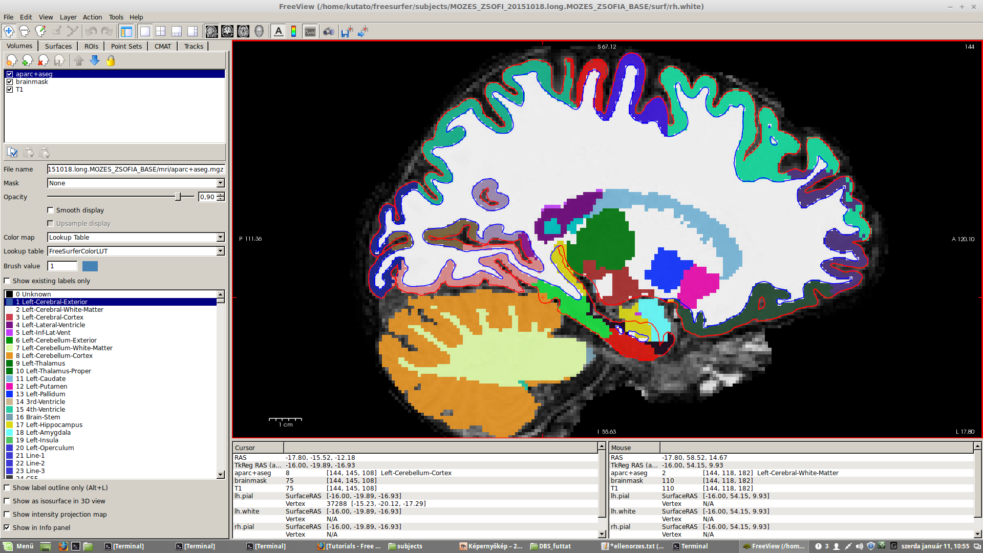
In some of our subjects the border of ctx_lh_parahippocampal (green color) is inconsistent with the border of pial surface (red color) in the sense that some small region within the pial surface is segmented as cerebellum (orange) coming from the subcortical segmentation stream. This problem is present in all three planes. It is hard to decide whether the colored border of ctx_lh_parahippocampal or the pial surface is more realistic, but it would be nice to know which one is used for the parahippocampal statistics in lh.aparc.stats file. Do I need to correct for this inaccuracy? How should I correct this type of missegmentation?
Best Regards, Gabor petzinger.gabor@gmail.com
freesurfer@nmr.mgh.harvard.edu
-
 Gabor Perlaki