
Hello,
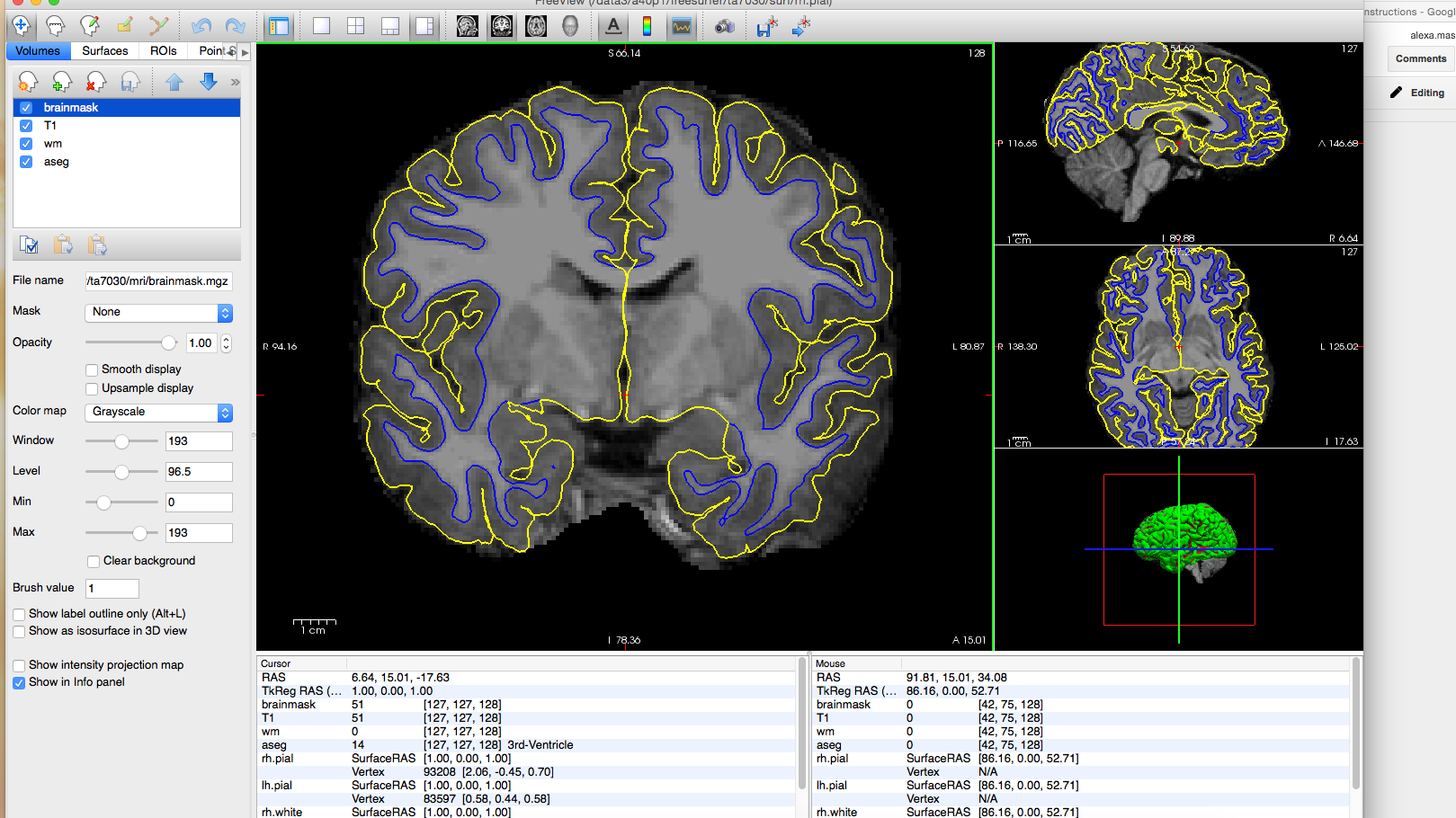
I attended the September 2016 Freesurfer training course, and I am seeking help to resolve a consistent issue with white and pial surface extraction that seems to be rooted in poor intensity normalization. Our lab is working with a pediatric dataset acquired on a 3T scanner and recon-all was run in Freesurfer v5.3. Throughout the dataset, we have found that the surfaces are cutting off white matter and excluding a significant amount of gray matter, particularly in the lateral temporal lobes. I have attached several images to illustrate the problem. As you can see in the images, the pial surface in the lateral temporal lobes hugs close to the white/gray boundary and excludes gray matter.
We have used the -3T flag when running recon-all for all subjects, and we have manually changed the smoothing distance in mi_nu_correct to 30 for several subjects in an attempt to improve this, but we did not get a better result. We are hesitant to address this problem using control points because it would require adding many control points across many slices in nearly all the subjects, and we would like to avoid the reliability/replicability issues of so much manual editing. (Not to mention the time required to do such heavy editing). Is there any way to address this automatically through the recon pipeline?
Many thanks,
Meaghan Perdue

Hi Meaghan
it looks like the white surface is pretty accurate but the gm doesn't get out far enough. I would play with some of the expert options to mris_make_surfaces like max_gray_at_csf_border. If you upload a subject I can try it out and see if I can improve things and get back to you.
cheers Bruce
On Wed, 22 Mar 2017, Meaghan Perdue wrote:
  Hello,

  I attended the September 2016 Freesurfer training course, and I am
seeking help to resolve a consistent issue with white and pial surface extraction that seems to be rooted in poor intensity normalization. Our lab is working with a pediatric dataset acquired on a 3T scanner and recon-all was run in Freesurfer v5.3. Throughout the dataset, we have found that the surfaces are cutting off white matter and excluding a significant amount of gray matter, particularly in the lateral temporal lobes. I have attached several images to illustrate the problem. As you can see in the images, the pial surface in the lateral temporal lobes hugs close to the white/gray boundary and excludes gray matter. We have used the -3T flag when running recon-all for all subjects, and we have manually changed the smoothing distance in mi_nu_correct to 30 for several subjects in an attempt to improve this, but we did not get a better result. We are hesitant to address this problem using control points because it would require adding many control points across many slices in nearly all the subjects, and we would like to avoid the reliability/replicability issues of so much manual editing. (Not to mention the time required to do such heavy editing). Is there any way to address this automatically through the recon pipeline? Many thanks, Meaghan Perdue
ta7029.zip https://drive.google.com/a/uconn.edu/file/d/0BzzXIfjzDH-kZFVzOEJqUTVmZVE/view?usp=drive_web Hi Bruce,
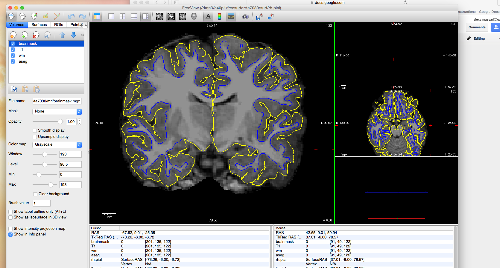
Thank you for your prompt response. I added an expert.opts file with the line "mris_make_surfaces -max_gray_at_csf_border 60 -max_csf 35" and ran recon-all -s ta7029 -autorecon2 -autorecon3 -expert ta7029/scripts/expert.opts -3T -qcache for one of the affected subjects, but it doesn't seem to have corrected the pial surface. I have attached the subject folder here. If you have time to take a look and try a couple of things, I would really appreciate it!
Thanks, Meaghan
On Wed, Mar 22, 2017 at 11:21 AM, Bruce Fischl fischl@nmr.mgh.harvard.edu wrote:
Hi Meaghan
it looks like the white surface is pretty accurate but the gm doesn't get out far enough. I would play with some of the expert options to mris_make_surfaces like max_gray_at_csf_border. If you upload a subject I can try it out and see if I can improve things and get back to you.
cheers Bruce
On Wed, 22 Mar 2017, Meaghan Perdue wrote:
Hello, I attended the September 2016 Freesurfer training course, and I amseeking help to resolve a consistent issue with white and pial surface extraction that seems to be rooted in poor intensity normalization. Our lab is working with a pediatric dataset acquired on a 3T scanner and recon-all was run in Freesurfer v5.3. Throughout the dataset, we have found that the surfaces are cutting off white matter and excluding a significant amount of gray matter, particularly in the lateral temporal lobes. I have attached several images to illustrate the problem. As you can see in the images, the pial surface in the lateral temporal lobes hugs close to the white/gray boundary and excludes gray matter.
We have used the -3T flag when running recon-all for all subjects,and we have manually changed the smoothing distance in mi_nu_correct to 30 for several subjects in an attempt to improve this, but we did not get a better result. We are hesitant to address this problem using control points because it would require adding many control points across many slices in nearly all the subjects, and we would like to avoid the reliability/replicability issues of so much manual editing. (Not to mention the time required to do such heavy editing). Is there any way to address this automatically through the recon pipeline?
  Many thanks,

  Meaghan Perdue
Freesurfer mailing list Freesurfer@nmr.mgh.harvard.edu https://mail.nmr.mgh.harvard.edu/mailman/listinfo/freesurfer
The information in this e-mail is intended only for the person to whom it is addressed. If you believe this e-mail was sent to you in error and the e-mail contains patient information, please contact the Partners Compliance HelpLine at http://www.partners.org/complianceline . If the e-mail was sent to you in error but does not contain patient information, please contact the sender and properly dispose of the e-mail.
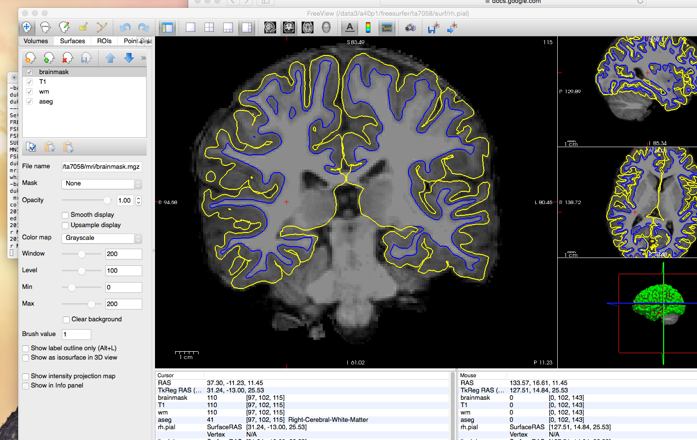
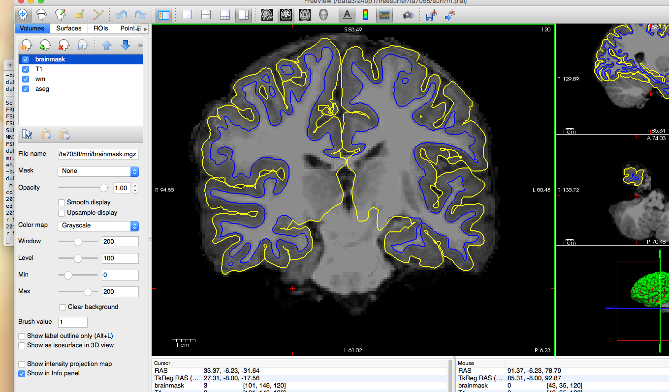
Hi Meaghan
I'm not sure that you reran the pial surface construction. In any case, I put a few control points in and added the following line to the expert opts file:
mris_make_surfaces -max_gray_at_csf_border 40 -max_csf 35
and got pretty good results. I would add a few more control points I think in spots where the white matter doesn't get segmented properly and/or the white surface doesn't include all the visible white matter (and the brain.mgz is significantly darker than 110).
I'll attach the control.data I created, which you can put in the subject's tmp dir and also the pial surface that I recreated.
Note this is using V6.
cheers Bruce
On Thu, 23 Mar 2017, Meaghan Perdue wrote:
[icon_10_generic_list.png] ta7029.zip[x_8px.png] Hi Bruce, Thank you for your prompt response. I added an expert.opts file with the line "mris_make_surfaces -max_gray_at_csf_border 60 -max_csf 35" and ran recon-all -s ta7029 -autorecon2 -autorecon3 -expert ta7029/scripts/expert.opts -3T -qcache for one of the affected subjects, but it doesn't seem to have corrected the pial surface. I have attached the subject folder here. If you have time to take a look and try a couple of things, I would really appreciate it!
Thanks, Meaghan
On Wed, Mar 22, 2017 at 11:21 AM, Bruce Fischl fischl@nmr.mgh.harvard.edu wrote: Hi Meaghan
  it looks like the white surface is pretty accurate but the gm doesn't get out far enough. I would play with some of the expert
  options to mris_make_surfaces like max_gray_at_csf_border. If you upload a subject I can try it out and see if I can improve things
  and get back to you.

  cheers
  Bruce


  On Wed, 22 Mar 2017, Meaghan Perdue wrote:


              Hello,

              I attended the September 2016 Freesurfer training course, and I am seeking help to resolve a consistent issue with
        white and pial surface extraction
              that seems to be rooted in poor intensity normalization. Our lab is working with a pediatric dataset acquired on a
        3T scanner and recon-all was run in
              Freesurfer v5.3. Throughout the dataset, we have found that the surfaces are cutting off white matter and
        excluding a significant amount of gray
              matter, particularly in the lateral temporal lobes.  I have attached several images to illustrate the problem. As
        you can see in the images, the pial
              surface in the lateral temporal lobes hugs close to the white/gray boundary and excludes gray matter.

              We have used the -3T flag when running recon-all for all subjects, and we have manually changed the smoothing
        distance in mi_nu_correct to 30 for
              several subjects in an attempt to improve this, but we did not get a better result. We are hesitant to address
        this problem using control points
              because it would require adding many control points across many slices in nearly all the subjects, and we would
        like to avoid the
              reliability/replicability issues of so much manual editing. (Not to mention the time required to do such heavy
        editing). Is there any way to address
              this automatically through the recon pipeline?


              Many thanks,

              Meaghan Perdue
Freesurfer mailing list Freesurfer@nmr.mgh.harvard.edu https://mail.nmr.mgh.harvard.edu/mailman/listinfo/freesurfer
The information in this e-mail is intended only for the person to whom it is addressed. If you believe this e-mail was sent to you in error and the e-mail contains patient information, please contact the Partners Compliance HelpLine at http://www.partners.org/complianceline . If the e-mail was sent to you in error but does not contain patient information, please contact the sender and properly dispose of the e-mail.
freesurfer@nmr.mgh.harvard.edu
-
 Bruce Fischl
 -
 Meaghan Perdue