
Hello,
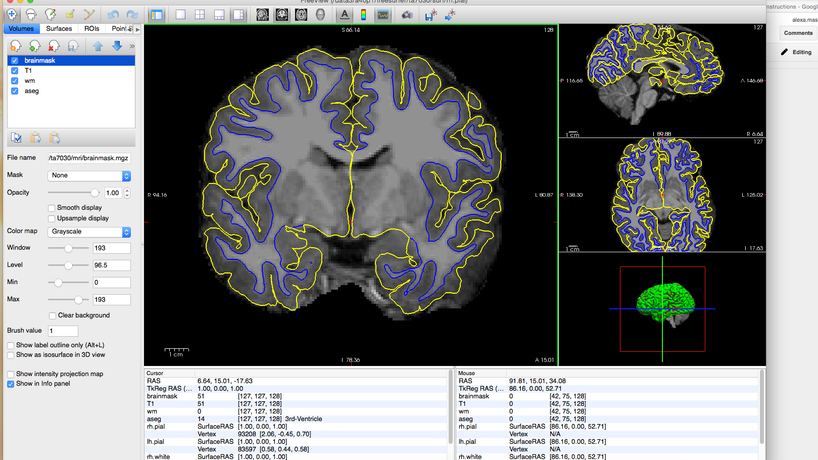
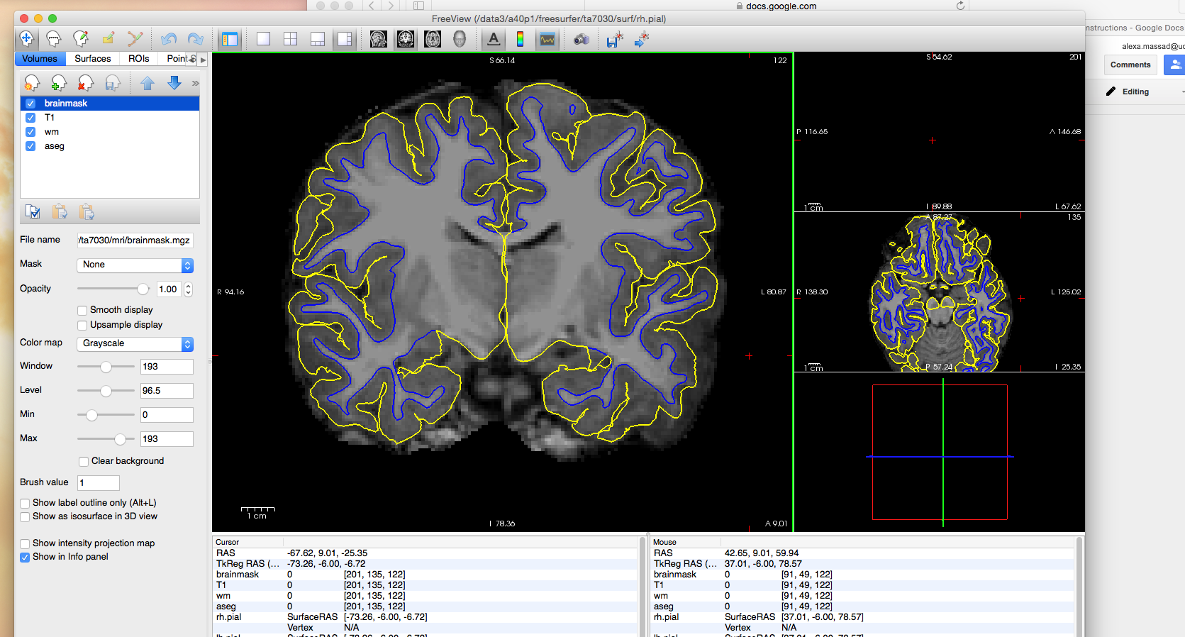
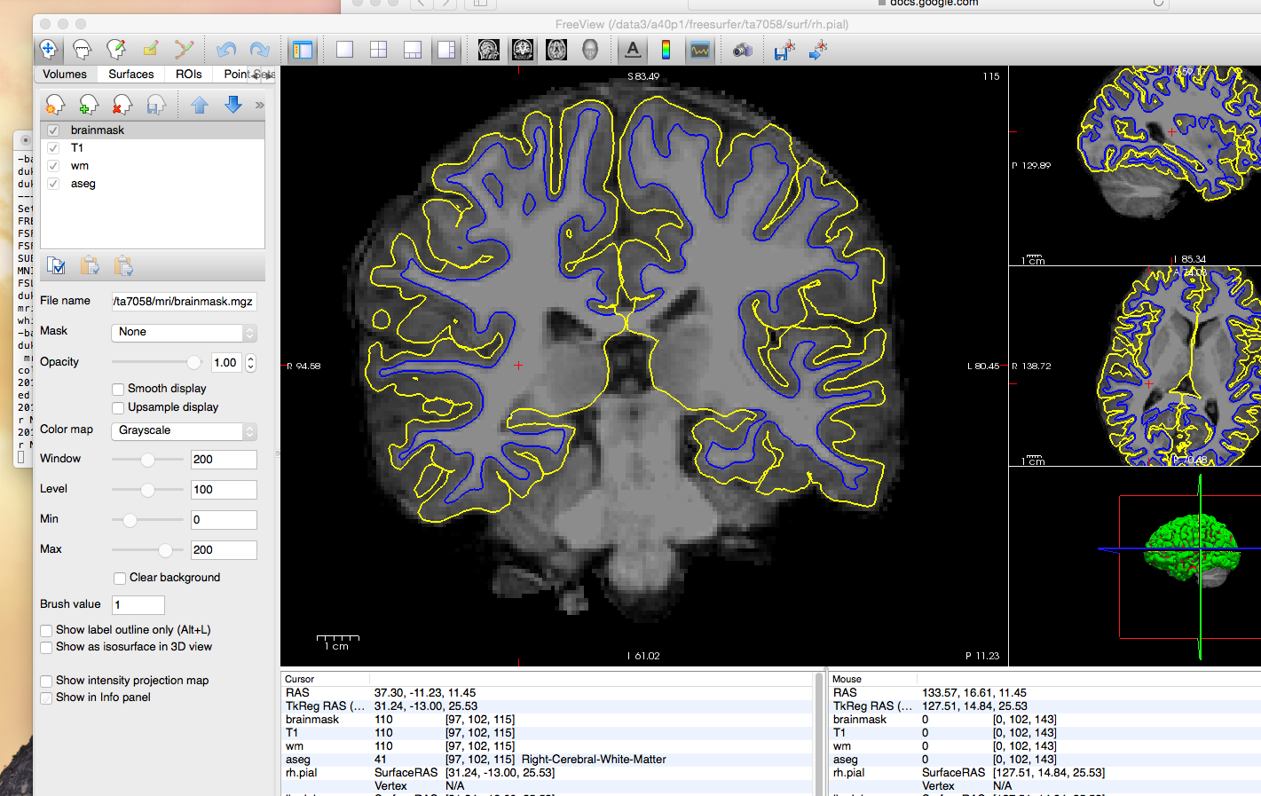
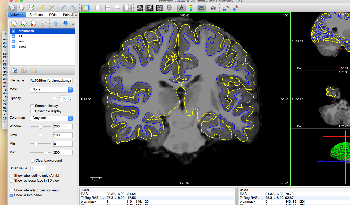
I attended the September 2016 Freesurfer training course, and I am seeking help to resolve a consistent issue with white and pial surface extraction that seems to be rooted in poor intensity normalization. Our lab is working with a pediatric dataset acquired on a 3T scanner and recon-all was run in Freesurfer v5.3. Throughout the dataset, we have found that the surfaces are cutting off white matter and excluding a significant amount of gray matter, particularly in the lateral temporal lobes. I have attached several images to illustrate the problem. As you can see in the images, the pial surface in the lateral temporal lobes hugs close to the white/gray boundary and excludes gray matter.
We have used the -3T flag when running recon-all for all subjects, and we have manually changed the smoothing distance in mi_nu_correct to 30 for several subjects in an attempt to improve this, but we did not get a better result. We are hesitant to address this problem using control points because it would require adding many control points across many slices in nearly all the subjects, and we would like to avoid the reliability/replicability issues of so much manual editing. (Not to mention the time required to do such heavy editing). Is there any way to address this automatically through the recon pipeline?
Many thanks,
Meaghan Perdue