
Hello Freesurfers,
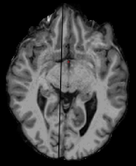
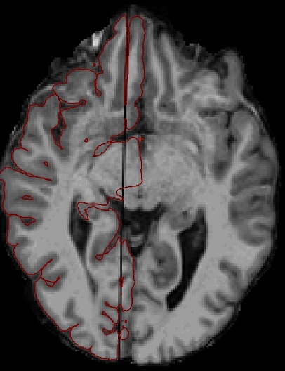
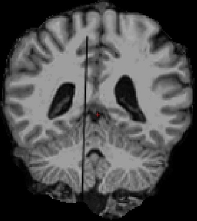
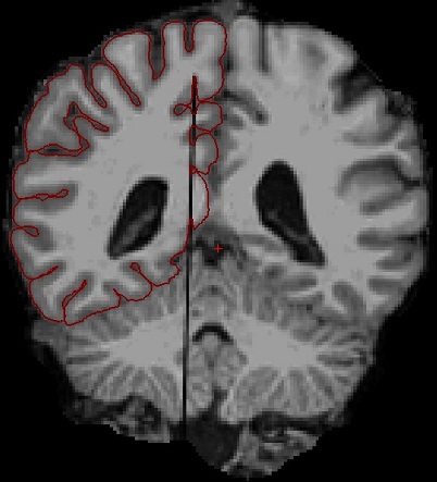
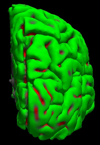
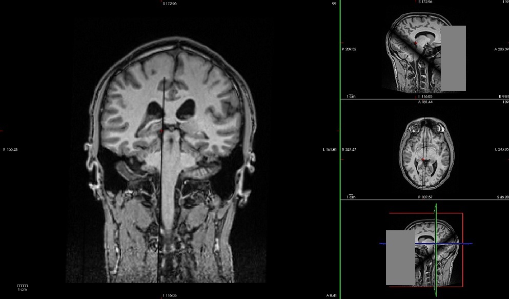
I have a subject who's missing part of one slice in the MR images (on the right hemisphere). This causes problems in the surface formation, as shown in the attached pictures.
First I tried to fix the problem by giving the central CC as seed point for the hemisphere cutting plane (-cc-crs), but this didn't help. The pictures were taken after I tried to fix the problem by filling in voxels manually into the wm.mgz. I managed to fix most of white surface problems, but the pial surface is still strongly drawn to the black line caused by the partly missing slice. This causes visible, canyon-like deformation as seen in the 3D view of pial surface.
The partly missing slice is probably due to an error in the scanning process - I looked at the DICOM images with a DICOM viewer, and the problem is also present there.
Do you have any suggestions on how to force the pial surface to follow a certain path, much like control points for wm? Or any other suggestions for fixing the problem? Getting the subject to do another MRI scan is unlikely at the moment.
Thank you in advance, Sami

Dear Sami
Instead of manual editing the FreeSurfer processed data (e.g. adding control points for white matter), one might try to manipulate the raw T1-w. image.
Maybe removing the incomplete slice and interpolating the signal of the neighboring slices into the missing slice help.
Then the surface model might be more or less accurately reconstructed and you can use it for analyses in other brain regions not affected by this missing slice.
I have never done this, but it might be worth trying.
Cheers Jürgen
On [DATE], "Sami Karadeniz" <[ADDRESS]> wrote:
Hello Freesurfers,
I have a subject who's missing part of one slice in the MR images (on the right hemisphere). This causes problems in the surface formation, as shown in the attached pictures.
First I tried to fix the problem by giving the central CC as seed point for the hemisphere cutting plane (-cc-crs), but this didn't help. The pictures were taken after I tried to fix the problem by filling in voxels manually into the wm.mgz. I managed to fix most of white surface problems, but the pial surface is still strongly drawn to the black line caused by the partly missing slice. This causes visible, canyon-like deformation as seen in the 3D view of pial surface.
The partly missing slice is probably due to an error in the scanning process - I looked at the DICOM images with a DICOM viewer, and the problem is also present there.
Do you have any suggestions on how to force the pial surface to follow a certain path, much like control points for wm? Or any other suggestions for fixing the problem? Getting the subject to do another MRI scan is unlikely at the moment.
Thank you in advance, Sami
Freesurfer mailing list Freesurfer@nmr.mgh.harvard.edu https://mail.nmr.mgh.harvard.edu/mailman/listinfo/freesurfer
The information in this e-mail is intended only for the person to whom it is addressed. If you believe this e-mail was sent to you in error and the e-mail contains patient information, please contact the Partners Compliance HelpLine at http://www.partners.org/complianceline . If the e-mail was sent to you in error but does not contain patient information, please contact the sender and properly dispose of the e-mail.
---------------------------------------------------------------------------- Jürgen Hänggi, Ph.D. Division Neuropsychology Institute of Psychology University of Zurich Binzmuehlestrasse 14, PO Box 25 8050 Zurich, Switzerland 0041 44 635 73 97 (phone office) 0041 76 445 86 84 (phone mobile) 0041 44 635 74 09 (fax office) BIN 4.D.04 (office room number) j.haenggi[at]psychologie.uzh.ch (email) http://www.psychologie.uzh.ch/neuropsy/ (website) http://www.juergenhaenggi.ch (private website)
This e-mail (and any attachment/s) contains confidential and/or privileged information. If you are not the intended recipient (or have received this e-mail in error) please notify the sender immediately and destroy this e-mail. Any unauthorised copying, disclosure or distribution of the material in this e-mail is strictly forbidden. ----------------------------------------------------------------------------

I second this approach. Much easier than including a blank slice Bruce On Thu, 5 Jun 2014, Jürgen Hänggi wrote:
Dear Sami
Instead of manual editing the FreeSurfer processed data (e.g. adding control points for white matter), one might try to manipulate the raw T1-w. image.
Maybe removing the incomplete slice and interpolating the signal of the neighboring slices into the missing slice help.
Then the surface model might be more or less accurately reconstructed and you can use it for analyses in other brain regions not affected by this missing slice.
I have never done this, but it might be worth trying.
Cheers Jürgen
On [DATE], "Sami Karadeniz" <[ADDRESS]> wrote:
Hello Freesurfers,
I have a subject who's missing part of one slice in the MR images (on the right hemisphere). This causes problems in the surface formation, as shown in the attached pictures.
First I tried to fix the problem by giving the central CC as seed point for the hemisphere cutting plane (-cc-crs), but this didn't help. The pictures were taken after I tried to fix the problem by filling in voxels manually into the wm.mgz. I managed to fix most of white surface problems, but the pial surface is still strongly drawn to the black line caused by the partly missing slice. This causes visible, canyon-like deformation as seen in the 3D view of pial surface.
The partly missing slice is probably due to an error in the scanning process - I looked at the DICOM images with a DICOM viewer, and the problem is also present there.
Do you have any suggestions on how to force the pial surface to follow a certain path, much like control points for wm? Or any other suggestions for fixing the problem? Getting the subject to do another MRI scan is unlikely at the moment.
Thank you in advance, Sami
Freesurfer mailing list Freesurfer@nmr.mgh.harvard.edu https://mail.nmr.mgh.harvard.edu/mailman/listinfo/freesurfer
The information in this e-mail is intended only for the person to whom it is addressed. If you believe this e-mail was sent to you in error and the e-mail contains patient information, please contact the Partners Compliance HelpLine at http://www.partners.org/complianceline . If the e-mail was sent to you in error but does not contain patient information, please contact the sender and properly dispose of the e-mail.
Jürgen Hänggi, Ph.D. Division Neuropsychology Institute of Psychology University of Zurich Binzmuehlestrasse 14, PO Box 25 8050 Zurich, Switzerland 0041 44 635 73 97 (phone office) 0041 76 445 86 84 (phone mobile) 0041 44 635 74 09 (fax office) BIN 4.D.04 (office room number) j.haenggi[at]psychologie.uzh.ch (email) http://www.psychologie.uzh.ch/neuropsy/ (website) http://www.juergenhaenggi.ch (private website)
This e-mail (and any attachment/s) contains confidential and/or privileged information. If you are not the intended recipient (or have received this e-mail in error) please notify the sender immediately and destroy this e-mail. Any unauthorised copying, disclosure or distribution of the material in this e-mail is strictly forbidden.
Freesurfer mailing list Freesurfer@nmr.mgh.harvard.edu https://mail.nmr.mgh.harvard.edu/mailman/listinfo/freesurfer
freesurfer@nmr.mgh.harvard.edu
-
 Bruce Fischl
 -
 Jürgen Hänggi
 -
 Sami Karadeniz