
Hello Freesurfers,
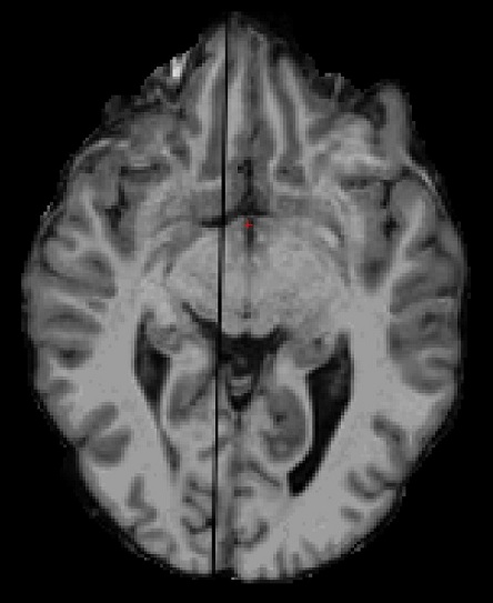
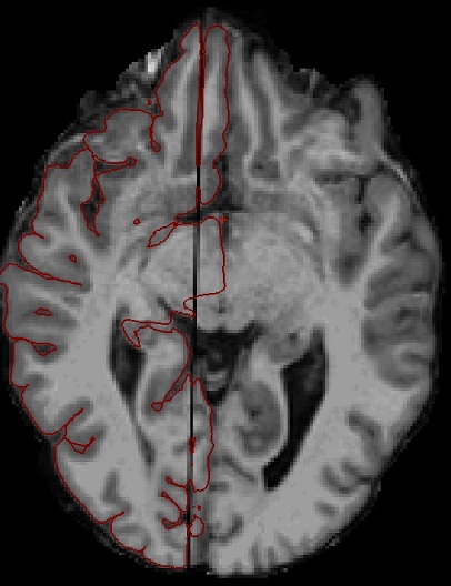
I have a subject who's missing part of one slice in the MR images (on the right hemisphere). This causes problems in the surface formation, as shown in the attached pictures.
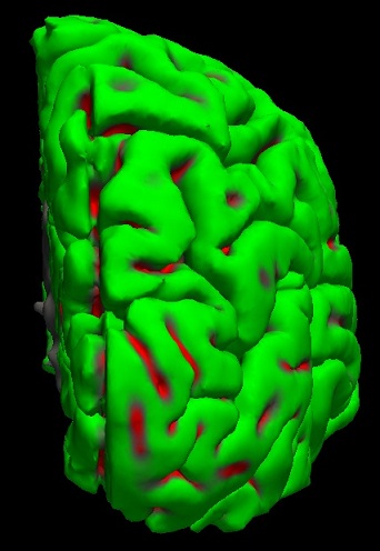
First I tried to fix the problem by giving the central CC as seed point for the hemisphere cutting plane (-cc-crs), but this didn't help. The pictures were taken after I tried to fix the problem by filling in voxels manually into the wm.mgz. I managed to fix most of white surface problems, but the pial surface is still strongly drawn to the black line caused by the partly missing slice. This causes visible, canyon-like deformation as seen in the 3D view of pial surface.
The partly missing slice is probably due to an error in the scanning process - I looked at the DICOM images with a DICOM viewer, and the problem is also present there.
Do you have any suggestions on how to force the pial surface to follow a certain path, much like control points for wm? Or any other suggestions for fixing the problem? Getting the subject to do another MRI scan is unlikely at the moment.
Thank you in advance, Sami