
Hi to all,
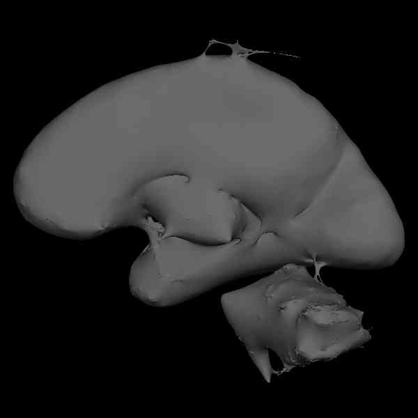
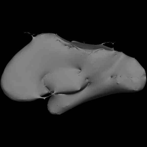
In our last analysis, among 12 subjects after "-stage 1" and "-stage 2" we got only 3 reasonable brain surfaces. Most of the other brains have a very odd shape: -pieces of skull -coharse lack of cortex -holes -bridges -bad cutting planes -cerebellar attachments It seems to be a segmentation problem. How to solve some of these problems is clearly explained in the 'FreesurferGuide' but the whole analysis seems to be "too much affected"!
I have to fix those errors manually? Can repeat the -stage 1 changing some parameters in the intensity normalization and/or segmentation process? I attach some examples (small jpg files)
Thank you in advance.
Cheers Francesco Musso
Laboratory of Molecular Neuroimaging & Electrophysiology, Department of Psychiatry, Johannes Gutenberg-University, Mainz, Germany

Hi Francesco,
these look pretty bad. Can you describe the acquisitions? How many? Did you motion correct and average them? What is the resolution? What field strength? What receive coil(s)?
cheers, Bruce
On Thu, 16 Sep 2004, F.Musso wrote:
Hi to all,
In our last analysis, among 12 subjects after "-stage 1" and "-stage 2" we got only 3 reasonable brain surfaces. Most of the other brains have a very odd shape: -pieces of skull -coharse lack of cortex -holes -bridges -bad cutting planes -cerebellar attachments It seems to be a segmentation problem. How to solve some of these problems is clearly explained in the 'FreesurferGuide' but the whole analysis seems to be "too much affected"!
I have to fix those errors manually? Can repeat the -stage 1 changing some parameters in the intensity normalization and/or segmentation process? I attach some examples (small jpg files)
Thank you in advance.
Cheers Francesco Musso
Laboratory of Molecular Neuroimaging & Electrophysiology, Department of Psychiatry, Johannes Gutenberg-University, Mainz, Germany
Hi Bruce and Nguyen,
Thanks for your reply. The scanner is a Sonata Siemens of 1.5 T with a new head coil -I'm waiting the feedback from the engineer of the neurorad for more technical details over this coil- The following is the output of 'mri_convert' over the native scanner data (mr.1.3.12.2.1107....) I cancel the subject name for a privacy issue :-) --------------------------------------------------------------------------- Identification NumarisVer syngo MR 2002B 4VA21A ScannerModel Sonata PatientName XXXXXXXXX Date and time StudyDate 20040630 StudyTime 182400.000000 SeriesTime 184547.703000 AcqTime 183813.397498 Acquisition parameters PulseSeq *tfl3d1_ns Protocol MPRAGE_sag_1.0mm_sense2 PhEncDir ROW EchoNo 0 FlipAngle 0.261799 EchoTime 3.93 InversionTime 1100 RepetitionTime 2160 PhEncFOV 256 ReadoutFOV 256 Image information RunNo 1 SeriesNo 2 ImageNo 1 NImageRows 512 NImageCols 512 NFrames 1 SliceArraylSize 1 IsMosaic 0 ImgPos 90.3748 165.9758 123.3529 VolRes 0.5000 0.5000 1.0000 VolDim 512 512 176 Vc -0.0000 -1.0000 0.0000 Vr -0.0000 -0.0000 -1.0000 Vs -1.0000 -0.0000 0.0000 VolCenter 2.3748 37.9758 -4.6471 TR=2160.00, TE=3.93, TI=1100.00, flip angle=15.00 i_ras = (-0, -1, 0) j_ras = (-0, -0, -1) k_ras = (-1, -0, 0) Original Data has (0.5, 0.5, 1) mm size and (512, 512, 176) voxels. Data is conformed to 1 mm size and 256 voxels for all directions changing data type from 4 to 0 (noscale = 0)... MRIchangeType: Building histogram Reslicing using trilinear interpolation writing to .... -----------------------------------------------------------------------
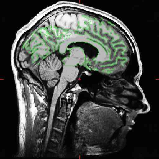
We used 2 acquisitions. The acquisitions were taked one at the beginning and one at the end of the scanning session [MPRAGE1-ODDBALL-STERNBERG-DTI-MPRAGE2] so after 1 hour +/-. The scanner perform a 'real time motion correction' over the scanning session. I use the 'mri_motion_correct' script to register and average the acquisitions, and the 'talairach2' script to obtain the trasform.xfm. Then I use the GUI for the -stage 1 (process volume) and -stage 2 (create surface). I attach an example of the segmentation.
----- Original Message ----- From: "Bruce Fischl" fischl@nmr.mgh.harvard.edu To: "F.Musso" musso@psychiatrie.klinik.uni-mainz.de Cc: freesurfer@nmr.mgh.harvard.edu Sent: Friday, September 17, 2004 4:08 AM Subject: Re: bad segmentation [newbie]
Hi Francesco,
these look pretty bad. Can you describe the acquisitions? How many? Did
you
motion correct and average them? What is the resolution? What field strength? What receive coil(s)?
cheers, Bruce
freesurfer@nmr.mgh.harvard.edu
-
 Bruce Fischl
 -
 F.Musso