
Hi Bruce and Nguyen,
Thanks for your reply. The scanner is a Sonata Siemens of 1.5 T with a new head coil -I'm waiting the feedback from the engineer of the neurorad for more technical details over this coil- The following is the output of 'mri_convert' over the native scanner data (mr.1.3.12.2.1107....) I cancel the subject name for a privacy issue :-) --------------------------------------------------------------------------- Identification NumarisVer syngo MR 2002B 4VA21A ScannerModel Sonata PatientName XXXXXXXXX Date and time StudyDate 20040630 StudyTime 182400.000000 SeriesTime 184547.703000 AcqTime 183813.397498 Acquisition parameters PulseSeq *tfl3d1_ns Protocol MPRAGE_sag_1.0mm_sense2 PhEncDir ROW EchoNo 0 FlipAngle 0.261799 EchoTime 3.93 InversionTime 1100 RepetitionTime 2160 PhEncFOV 256 ReadoutFOV 256 Image information RunNo 1 SeriesNo 2 ImageNo 1 NImageRows 512 NImageCols 512 NFrames 1 SliceArraylSize 1 IsMosaic 0 ImgPos 90.3748 165.9758 123.3529 VolRes 0.5000 0.5000 1.0000 VolDim 512 512 176 Vc -0.0000 -1.0000 0.0000 Vr -0.0000 -0.0000 -1.0000 Vs -1.0000 -0.0000 0.0000 VolCenter 2.3748 37.9758 -4.6471 TR=2160.00, TE=3.93, TI=1100.00, flip angle=15.00 i_ras = (-0, -1, 0) j_ras = (-0, -0, -1) k_ras = (-1, -0, 0) Original Data has (0.5, 0.5, 1) mm size and (512, 512, 176) voxels. Data is conformed to 1 mm size and 256 voxels for all directions changing data type from 4 to 0 (noscale = 0)... MRIchangeType: Building histogram Reslicing using trilinear interpolation writing to .... -----------------------------------------------------------------------
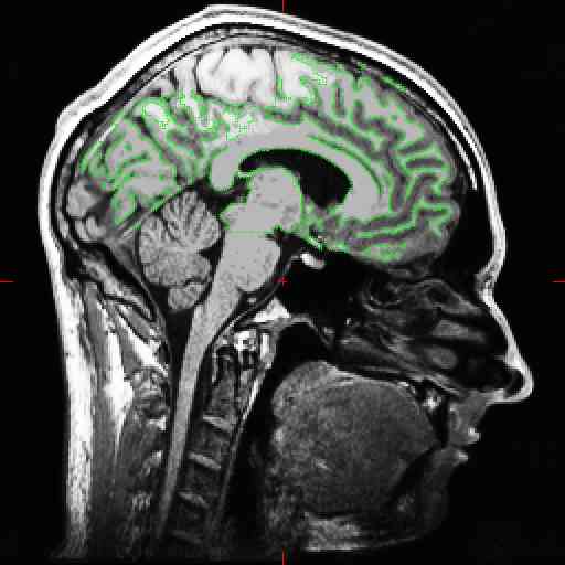
We used 2 acquisitions. The acquisitions were taked one at the beginning and one at the end of the scanning session [MPRAGE1-ODDBALL-STERNBERG-DTI-MPRAGE2] so after 1 hour +/-. The scanner perform a 'real time motion correction' over the scanning session. I use the 'mri_motion_correct' script to register and average the acquisitions, and the 'talairach2' script to obtain the trasform.xfm. Then I use the GUI for the -stage 1 (process volume) and -stage 2 (create surface). I attach an example of the segmentation.
----- Original Message ----- From: "Bruce Fischl" fischl@nmr.mgh.harvard.edu To: "F.Musso" musso@psychiatrie.klinik.uni-mainz.de Cc: freesurfer@nmr.mgh.harvard.edu Sent: Friday, September 17, 2004 4:08 AM Subject: Re: bad segmentation [newbie]
Hi Francesco,
these look pretty bad. Can you describe the acquisitions? How many? Did
you
motion correct and average them? What is the resolution? What field strength? What receive coil(s)?
cheers, Bruce