Re: [Freesurfer] the pial and wm are larger than the inflated (beyond the inflated image)


Hi Star
this isn't a problem. The inflated has 1-to-1 correspondence with both the pial and the white. The position in space of the inflated relative to them is irrelevant. If you want to assess accuracy you can overlay the white and pial on the MRI volumes and look at that in slices
cheers Bruce
On Thu, 3 Jan 2019, xi star wrote:
External Email - Use Caution
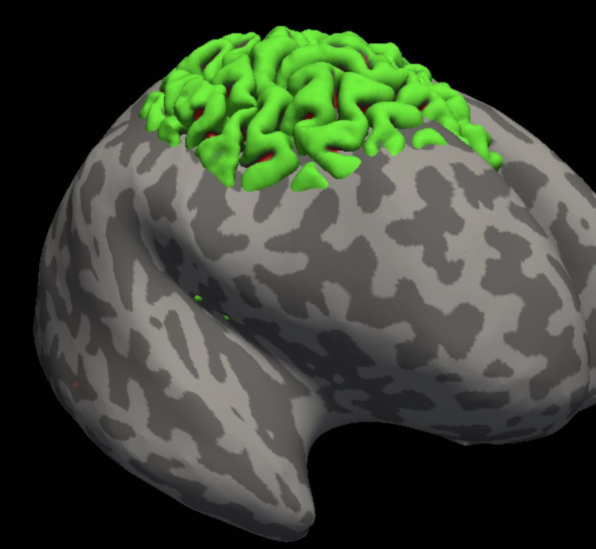
I used freeview to visualized the inflated surface. The image is in the attachment
席思达12级临床医学(8 年制) 复旦大学上海医学院
On 01/03/2019 01:14, Bruce Fischl wrote: Hi Star
  what do you mean by "outside the inflated surfaces"? Can you
  send an
  image? The inflated surface should not be viewed overlaid on a
  slice as
  it no longer corresponds directly to the underlying MRI. You
  should
  visualize the ?h.white and ?h.pial surfaces instead to assess
  accuracy

  cheers
  Bruce
  On
  Thu, 3 Jan 2019, xi star wrote:

  >
  >         External Email - Use Caution
  >
  >       I'm a beginner on fs. my data was done with no error in
  all three
  > parts of recon-all (autorecon1 2 & 3), and I've checked the
  surfaces each
  > time.
  >       However, I found the pial and wm of part of the frontal
  and parietal
  > lobel was outside the inflated surfaces. As you can see in the
  attachment.
  >       I wonder what's wrong with the data or how i can find
  and fix the
  > problem. I really puzzled and want your help
  > Best wish,
  > Star
  >
  >
  >
  >_______________________________________________
  Freesurfer mailing list
  Freesurfer@nmr.mgh.harvard.edu
https://mail.nmr.mgh.harvard.edu/mailman/listinfo/freesurfer_______________ ________________________________ Freesurfer mailing list Freesurfer@nmr.mgh.harvard.edu https://mail.nmr.mgh.harvard.edu/mailman/listinfo/freesurfer
freesurfer@nmr.mgh.harvard.edu
-
 Bruce Fischl
 -
 xi star