
Dear all,
I've two questions which I couldn't get answered in your wiki tutorials. I hope you can help me.
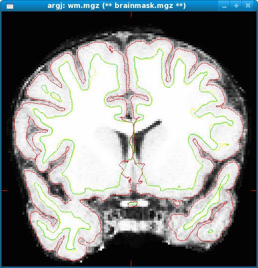
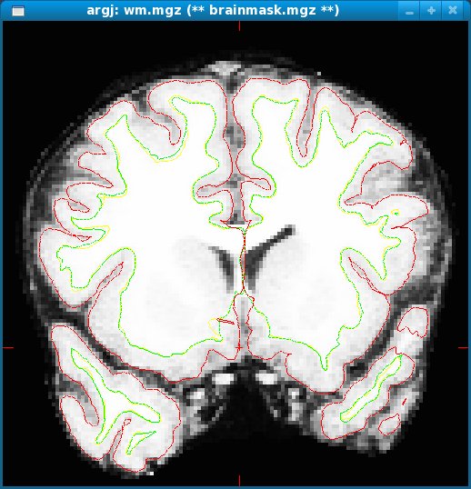
- I have some processed images that have in the medial part of the brain intersection of the pial matter and the grey matter (see image in attachment). From the tutorial I could only learn about removing dura segmented as pial matter and filling in wm on lesions/ventricles. In my case if I delete voxels I'll loose wm/gm information is there anything I can do?
- Also, there are cases where the pial matter does not cover the full gm, particularly at the base of the skull and at the temporal lobes (see attachment). What do you recommend me to do?
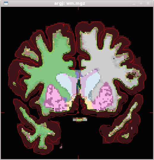
One thing though is that the annotation seems to be ok (see attachs). So do I really have to correct for the pial and wm surface, as I'm asking above, as I'm interested in studying both cortical thicknesses and subcortical volumes of gm structures, wm and ventricles, or do not have to worry about it?
Regarding QDEC, when preprocessing/preparing images for analysis do we have to have all the fsaverage folder together with all the subjects (lets say patients and controls), or can I have the fsaverage folder in two or more subfolders were I have segregated patient subjects and control subjects? This is to ask you also, do the fsaverage folder is changed by the qdec -cache processing or is it used only as a template for each subject (patient and control), and data is only created in patient/control individual subject folders?
Many thanks and best regards, Hugo Ferreira
freesurfer@nmr.mgh.harvard.edu
-
 Hugo Ferreira