
External Email - Use Caution
Hello FreeSurfer Developers,
We are completing quality control in FreeView of two longitudinal datasets (participants age: 9-25) and have noticed that in one dataset the brains look much more 'spiky' than in the other dataset. More than half of the brains have some 'spikes', < 10% has severe spikes; the most obvious ones correspond with both underestimations and overestimations in frontal regions of the brain (e.g., frontal pole, rostral middle frontal and superior frontal). The datasets are collected on two different scanners:
Dataset with the spiky brains:
* Structural MRI data were acquired using a Philips Achieva 3.0 Tesla MRI scanner with a standard whole-head coil. The following parameters were used: FOV = 224 mm x 178.5 mm x 168 mm; TR = 9.8 ms; TE = 4.6 ms; flip angle = 8 degrees; voxel size = 0.875 mm x 0.875 mm x 0.875 mm. The duration of the anatomical scan was 297.8 s. To reduce head motion, foam inserts were used and a film was projected on a screen.
Dataset without spiky brains:
* Structural MRI data were acquired using a Philips Achieva 3.0 TX MRI scanner with a standard whole-head coil. The following parameters were used: FOV = 250 mm × 196 mm × 170 mm; TR = 7.9 ms; TE = 3.5 ms, flip angle = 8 degrees, 228 × 177 × 155 slices; voxel size = 1.1 × 1.1 × 1.1 mm. The duration of the anatomical scan was 251 s. To reduce head motion, foam inserts were used.
We are not too sure what to do about these. Manual skull stripping, adjusting the watershed threshold and using control points are not perfect solutions for various reasons. And we have noticed that the spikiness is reduced in some brains when the scans are processed in version 6.0 compared to version 7.3 - but otherwise version 7.3 seems like the better fit.
We couldn't find anything in the archives, but we wonder - have you come across this before? Do you have another idea for us to try? Or is it possible that data from these regions would be (mostly) unimpacted by the appearance of small, infrequent spikes? If we want to use global brain measures, do you think Freesurfer gives a reliable estimation or would you advise us to exclude them from the analyses due to bad data quality.
We have attached one picture of severe spikes so you know what we mean. We have started to rate the spikes from 1 (small/infrequent) to 4 (large/frequent).
Thank you in advance for any ideas you can share with us.
Best wishes,
Manon Marieke

This sometimes happens with noisy data. The difference in SNR between those two scans is a factor of 2. What is your recon-all command line? Can you send pics of the brain image with the surface overlaid on a slice with one of the spikes? What does the white surface look like in those regions? Is it actually grabbing part of the skull or dura?
On 12/12/2024 4:44 AM, Bos, M.G.N. (Marieke) wrote:
External Email - Use Caution
Hello FreeSurfer Developers,
We are completing quality control in FreeView of two longitudinal datasets (participants age: 9-25) and have noticed that in one dataset the brains look much more 'spiky' than in the other dataset. More than half of the brains have some 'spikes', < 10% has severe spikes; the most obvious ones correspond with both underestimations and overestimations in frontal regions of the brain (e.g., frontal pole, rostral middle frontal and superior frontal). The datasets are collected on two different scanners:
Dataset with the spiky brains:
- Structural MRI data were acquired using a Philips Achieva 3.0 Tesla MRI scanner with a standard whole-head coil. The following parameters were used: FOV = 224 mm x 178.5 mm x 168 mm; TR = 9.8 ms; TE = 4.6 ms; flip angle = 8 degrees; voxel size = 0.875 mm x 0.875 mm x 0.875 mm. The duration of the anatomical scan was 297.8 s. To reduce head motion, foam inserts were used and a film was projected on a screen.
Dataset without spiky brains:
- Structural MRI data were acquired using a Philips Achieva 3.0 TX MRI scanner with a standard whole-head coil. The following parameters were used: FOV = 250 mm × 196 mm × 170 mm; TR = 7.9 ms; TE = 3.5 ms, flip angle = 8 degrees, 228 × 177 × 155 slices; voxel size = 1.1 × 1.1 × 1.1 mm. The duration of the anatomical scan was 251 s. To reduce head motion, foam inserts were used.
We are not too sure what to do about these. Manual skull stripping, adjusting the watershed threshold and using control points are not perfect solutions for various reasons. And we have noticed that the spikiness is reduced in some brains when the scans are processed in version 6.0 compared to version 7.3 - but otherwise version 7.3 seems like the better fit.
We couldn't find anything in the archives, but we wonder - have you come across this before? Do you have another idea for us to try?
Or is it possible that data from these regions would be (mostly) unimpacted by the appearance of small, infrequent spikes? If we want to use global brain measures, do you think Freesurfer gives a reliable estimation or would you advise us to exclude them from the analyses due to bad data quality.
We have attached one picture of severe spikes so you know what we mean. We have started to rate the spikes from 1 (small/infrequent) to 4 (large/frequent).
Thank you in advance for any ideas you can share with us.
Best wishes,
Manon
Marieke
Freesurfer mailing list Freesurfer@nmr.mgh.harvard.edu https://mail.nmr.mgh.harvard.edu/mailman/listinfo/freesurfer
External Email - Use Caution
Thank you for your response, Douglas!
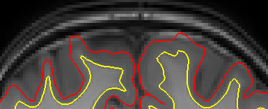
We have attached a picture of the pial surface over the orig volume where there is a severe spike. There are underestimations and overestimations - which seem to include dura - but there aren't always both.
Here is how recon-all appears in our bash scripts:
Recon-all Cross-sectional
while IFS= read -r file; do if [ -f "$input_dir/$file" ]; then # Extract timepoint and participant information from the file name filename=$(basename "$file") timepoint="${filename:1:1}" participant="${filename:3:4}"
# Execute recon-all command "$freesurfer_bin/recon-all" \ -i "${input_dir}${file}" \ -subjid "Z${timepoint}P${participant}" \ -sd "$output_dir" \ -all fi done < "$filename"
Recon-all Base Template
# Loop through each line in the file while IFS= read -r folder_name || [[ -n "$folder_name" ]]; do # Check if folder exists in input directory folder_path="$input_dir$folder_name" if [ -d "$folder_path" ]; then # Get participant ID participant="${folder_name:3}"
# Check if the participant has been processed if [ -n "${processed_participants[$participant]}" ]; then echo "Participant ${participant} already processed. Skipping." continue fi
# Construct -tp options for all time points tp_options="" for tp_folder in "${input_dir}"Z*P"${participant}"/; do if [ -d "$tp_folder" ]; then tp=$(basename "$tp_folder" | sed -n 's/Z([0-9]+)P'"${participant}"'/\1/p') if [ -n "$tp" ]; then tp_options+=" -tp Z${tp}P${participant}" fi fi done
# Run recon-all command $freesurfer_bin/recon-all \ -base "P${participant}Base" \ ${tp_options} \ -sd "${output_dir}" \ -all
# Mark participant as processed processed_participants[$participant]=1 fi done < "$filename"
Recon-all Longitudinal
# Loop through cross files for cross_file in "${cross_files[@]}"; do # Extract timepoint cross_folder_name=$(basename "$cross_file") cross_timepoint="${cross_folder_name:1:1}" cross_participant="${cross_folder_name:3}"
# Loop through base files for base_file in "${base_files[@]}"; do base_folder_name=$(basename "$base_file") base_participant="${base_folder_name:1:4}" if [ "$cross_participant" == "$base_participant" ]; then # Run command $freesurfer_bin/recon-all \ -long "${cross_file}" "${base_file}" \ -sd "${output_dir}" \ -all fi done done
Best wishes, Marieke & Manon ________________________________
From: freesurfer-bounces@nmr.mgh.harvard.edu freesurfer-bounces@nmr.mgh.harvard.edu On Behalf Of Douglas N. Greve Sent: donderdag 12 december 2024 14:40 To: freesurfer@nmr.mgh.harvard.edu Subject: Re: [Freesurfer] Spiky Brains
This sometimes happens with noisy data. The difference in SNR between those two scans is a factor of 2. What is your recon-all command line? Can you send pics of the brain image with the surface overlaid on a slice with one of the spikes? What does the white surface look like in those regions? Is it actually grabbing part of the skull or dura?
On 12/12/2024 4:44 AM, Bos, M.G.N. (Marieke) wrote:
External Email - Use Caution Hello FreeSurfer Developers,
We are completing quality control in FreeView of two longitudinal datasets (participants age: 9-25) and have noticed that in one dataset the brains look much more 'spiky' than in the other dataset. More than half of the brains have some 'spikes', < 10% has severe spikes; the most obvious ones correspond with both underestimations and overestimations in frontal regions of the brain (e.g., frontal pole, rostral middle frontal and superior frontal). The datasets are collected on two different scanners:
Dataset with the spiky brains:
1. Structural MRI data were acquired using a Philips Achieva 3.0 Tesla MRI scanner with a standard whole-head coil. The following parameters were used: FOV = 224 mm x 178.5 mm x 168 mm; TR = 9.8 ms; TE = 4.6 ms; flip angle = 8 degrees; voxel size = 0.875 mm x 0.875 mm x 0.875 mm. The duration of the anatomical scan was 297.8 s. To reduce head motion, foam inserts were used and a film was projected on a screen.
Dataset without spiky brains:
1. Structural MRI data were acquired using a Philips Achieva 3.0 TX MRI scanner with a standard whole-head coil. The following parameters were used: FOV = 250 mm × 196 mm × 170 mm; TR = 7.9 ms; TE = 3.5 ms, flip angle = 8 degrees, 228 × 177 × 155 slices; voxel size = 1.1 × 1.1 × 1.1 mm. The duration of the anatomical scan was 251 s. To reduce head motion, foam inserts were used.
We are not too sure what to do about these. Manual skull stripping, adjusting the watershed threshold and using control points are not perfect solutions for various reasons. And we have noticed that the spikiness is reduced in some brains when the scans are processed in version 6.0 compared to version 7.3 - but otherwise version 7.3 seems like the better fit.
We couldn't find anything in the archives, but we wonder - have you come across this before? Do you have another idea for us to try? Or is it possible that data from these regions would be (mostly) unimpacted by the appearance of small, infrequent spikes? If we want to use global brain measures, do you think Freesurfer gives a reliable estimation or would you advise us to exclude them from the analyses due to bad data quality.
We have attached one picture of severe spikes so you know what we mean. We have started to rate the spikes from 1 (small/infrequent) to 4 (large/frequent).
Thank you in advance for any ideas you can share with us.
Best wishes,
Manon Marieke
_______________________________________________
Freesurfer mailing list
Freesurfer@nmr.mgh.harvard.edumailto:Freesurfer@nmr.mgh.harvard.edu
https://secure-web.cisco.com/1UgBShKer5QYnMDxCoUDo8VEL5LbhpktKPGEpSRwDrEMOBx...
freesurfer@nmr.mgh.harvard.edu
-
 Bos, M.G.N. (Marieke)
 -
 Douglas N. Greve