
Hi -
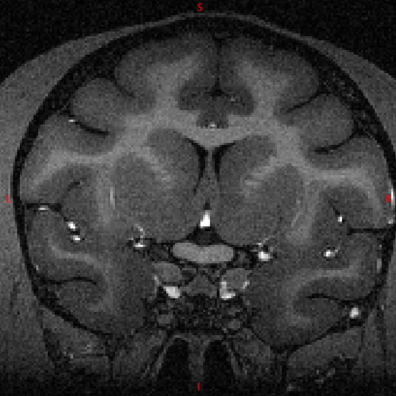
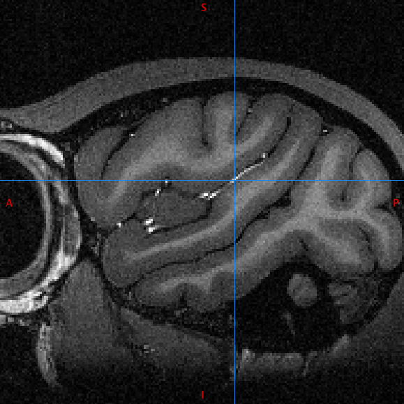
I wonder if anyone has experience in dealing with T1 images where there is hyperintenisty of blood vessels?
the data involved is MDEFT scans where only a head coil is used. I read it is a long solved problem for acquisition, but these scans do not correct it and nor will future scans.
the difficulty is getting 3 classes (CSF,WM,GM) for nu_correct,mri_normalise or FSL Fast or whatever.
It's quite a big obstacle for data that is otherwise quite excellent.
One would think one could just threshold it out after brain extraction and to an extent one can but it's very imperfect. segmenting 4 classes doesn't especially work either (so far)
if anyone has experience segmenting this stuff (especially getting mri_normalise to work) I'd be grateful for advice.
e.g. attached
Colin Reveley

Hi Colin,
On Oct 24, 2013, at 00:24 , Colin Reveley cmr25@sussex.ac.uk wrote:
Hi -
I wonder if anyone has experience in dealing with T1 images where there is hyperintenisty of blood vessels?
This looks like receive & transmit surface coil data to me. For MPRAGEs, in my experience, switching to receive only surface coil (and assuming a whole body scanner) and the body coil for transmit will darken the the blood vessels again. Or you could use 8mb/kg bodyweight feraheme to reduce the blood signal as well. Both methods will obviously not work retrospectively. I have no experience with MDEFT, so the fereheme might not work. (But if you go that route: feraheme is an excellent functional contrast agent in NHPs for EPIs)
the data involved is MDEFT scans where only a head coil is used. I read it is a long solved problem for acquisition, but these scans do not correct it and nor will future scans.
the difficulty is getting 3 classes (CSF,WM,GM) for nu_correct,mri_normalise or FSL Fast or whatever.
It's quite a big obstacle for data that is otherwise quite excellent.
One would think one could just threshold it out after brain extraction and to an extent one can but it's very imperfect. segmenting 4 classes doesn't especially work either (so far)
if anyone has experience segmenting this stuff (especially getting mri_normalise to work) I'd be grateful for advice.
How does it affect mri_normalize? I typically used: mri_normalize -n 1 -no1d -gentle ${src_dir}/${src_vol} ${targ_dir}/${targ_vol} -v -mprage on NHPs and: mri_normalize -f ${controlpoints} ${mri_normalize_options_string} -MASK ${src_dir}/${mask} ${src_dir}/${in_vol} ${targ_dir}/${unmasked_brain} -mprage for control point based normalization (which does not look required for your nice data) I guess for MDEFT I would skip -mprage.
I hope that helps at least some.
best Sebastian
e.g. attached
Colin Reveley <CORONAL_BLOOD.png><SAGG_BLOOD.png>_______________________________________________ Freesurfer mailing list Freesurfer@nmr.mgh.harvard.edu https://mail.nmr.mgh.harvard.edu/mailman/listinfo/freesurfer
The information in this e-mail is intended only for the person to whom it is addressed. If you believe this e-mail was sent to you in error and the e-mail contains patient information, please contact the Partners Compliance HelpLine at http://www.partners.org/complianceline . If the e-mail was sent to you in error but does not contain patient information, please contact the sender and properly dispose of the e-mail.
freesurfer@nmr.mgh.harvard.edu
-
 Colin Reveley
 -
 Sebastian Moeller