
Hi -
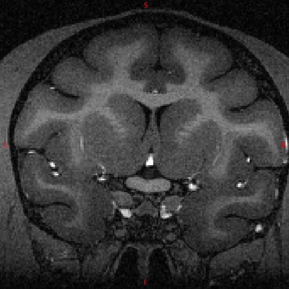
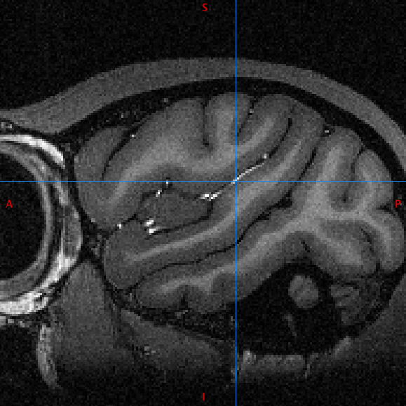
I wonder if anyone has experience in dealing with T1 images where there is hyperintenisty of blood vessels?
the data involved is MDEFT scans where only a head coil is used. I read it is a long solved problem for acquisition, but these scans do not correct it and nor will future scans.
the difficulty is getting 3 classes (CSF,WM,GM) for nu_correct,mri_normalise or FSL Fast or whatever.
It's quite a big obstacle for data that is otherwise quite excellent.
One would think one could just threshold it out after brain extraction and to an extent one can but it's very imperfect. segmenting 4 classes doesn't especially work either (so far)
if anyone has experience segmenting this stuff (especially getting mri_normalise to work) I'd be grateful for advice.
e.g. attached
Colin Reveley