
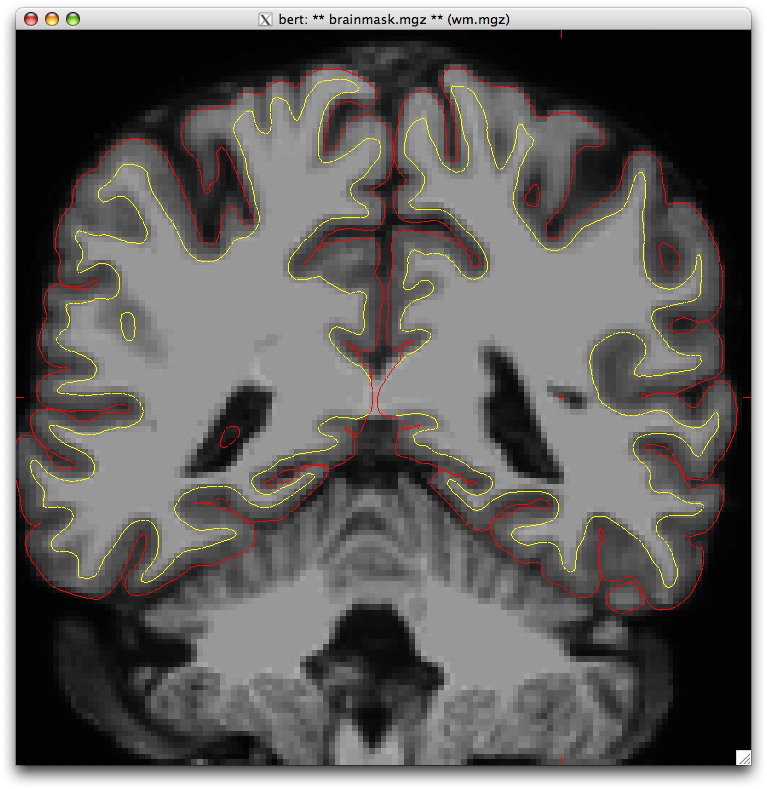
Hi, I was looking at some of my data and noticed that the white and pial surfaces occasionally invade the lateral ventricle (see attached picture). I went over to the bert sample dataset and noticed the same thing (coronal slice 87). Is this something I should be correcting or is it something that is ignored during the registration and labeling process? If I don't care about the choroid plexus volume, should I fill the white matter mask throughout the ventricle?
Thank you in advance, Mira
Mira Michelle Raman Scientific Programmer Center for Interdisciplinary Brain Sciences Research Stanford University 401 Quarry Rd. Stanford, CA 940305 http://cibsr.stanford.edu

Hi Mira,
usually the aseg will fill in the ventricle and choroid. What version are you using? What is the label of the stuff in the ventricle that wasn't filled?
cheers, Bruce On Mon, 17 Aug 2009, Mira Michelle Raman wrote:
Hi, I was looking at some of my data and noticed that the white and pial surfaces occasionally invade the lateral ventricle (see attached picture). I went over to the bert sample dataset and noticed the same thing (coronal slice 87). Is this something I should be correcting or is it something that is ignored during the registration and labeling process? If I don't care about the choroid plexus volume, should I fill the white matter mask throughout the ventricle?
Thank you in advance, Mira
Mira Michelle Raman Scientific Programmer Center for Interdisciplinary Brain Sciences Research Stanford University 401 Quarry Rd. Stanford, CA 940305 http://cibsr.stanford.edu

I've seen this in cases where the wm.mgz was created with an older version of freesurfer and then the subjects was re-run with a newer version. You might try deleting (or renaming!) the wm.mgz and re-running
recon-all -s 000502083470__0002 -autorecon2-cp
to see if the problem goes away.
doug
Bruce Fischl wrote:
Hi Mira,
usually the aseg will fill in the ventricle and choroid. What version are you using? What is the label of the stuff in the ventricle that wasn't filled?
cheers, Bruce On Mon, 17 Aug 2009, Mira Michelle Raman wrote:
Hi, I was looking at some of my data and noticed that the white and pial surfaces occasionally invade the lateral ventricle (see attached picture). I went over to the bert sample dataset and noticed the same thing (coronal slice 87). Is this something I should be correcting or is it something that is ignored during the registration and labeling process? If I don't care about the choroid plexus volume, should I fill the white matter mask throughout the ventricle?
Thank you in advance, Mira
Mira Michelle Raman Scientific Programmer Center for Interdisciplinary Brain Sciences Research Stanford University 401 Quarry Rd. Stanford, CA 940305 http://cibsr.stanford.edu
Freesurfer mailing list Freesurfer@nmr.mgh.harvard.edu https://mail.nmr.mgh.harvard.edu/mailman/listinfo/freesurfer
Hi Doug, That's a good thought, but we ran it through straight from the DICOM's both times 4.3 and 4.5 (obviously with different freesurfer subject IDs). So this couldn't be the problem, if it even is a problem. -Mira
----- Original Message ----- From: "Douglas N Greve" greve@nmr.mgh.harvard.edu To: "Bruce Fischl" fischl@nmr.mgh.harvard.edu Cc: "Mira Michelle Raman" mraman@stanford.edu, freesurfer@nmr.mgh.harvard.edu Sent: Monday, August 17, 2009 10:40:25 AM GMT -08:00 US/Canada Pacific Subject: Re: [Freesurfer] white/pial surface in lateral ventricle
I've seen this in cases where the wm.mgz was created with an older version of freesurfer and then the subjects was re-run with a newer version. You might try deleting (or renaming!) the wm.mgz and re-running
recon-all -s 000502083470__0002 -autorecon2-cp
to see if the problem goes away.
doug
Bruce Fischl wrote:
Hi Mira,
usually the aseg will fill in the ventricle and choroid. What version are you using? What is the label of the stuff in the ventricle that wasn't filled?
cheers, Bruce On Mon, 17 Aug 2009, Mira Michelle Raman wrote:
Hi, I was looking at some of my data and noticed that the white and pial surfaces occasionally invade the lateral ventricle (see attached picture). I went over to the bert sample dataset and noticed the same thing (coronal slice 87). Is this something I should be correcting or is it something that is ignored during the registration and labeling process? If I don't care about the choroid plexus volume, should I fill the white matter mask throughout the ventricle?
Thank you in advance, Mira
Mira Michelle Raman Scientific Programmer Center for Interdisciplinary Brain Sciences Research Stanford University 401 Quarry Rd. Stanford, CA 940305 http://cibsr.stanford.edu
Freesurfer mailing list Freesurfer@nmr.mgh.harvard.edu https://mail.nmr.mgh.harvard.edu/mailman/listinfo/freesurfer
freesurfer@nmr.mgh.harvard.edu
-
 Bruce Fischl
 -
 Douglas N Greve
 -
 Mira Michelle Raman