
17 Aug
2009
17 Aug
'09
1:04 p.m.
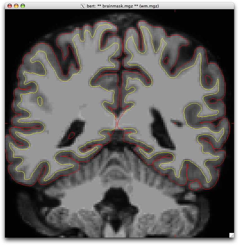
Hi, I was looking at some of my data and noticed that the white and pial surfaces occasionally invade the lateral ventricle (see attached picture). I went over to the bert sample dataset and noticed the same thing (coronal slice 87). Is this something I should be correcting or is it something that is ignored during the registration and labeling process? If I don't care about the choroid plexus volume, should I fill the white matter mask throughout the ventricle?
Thank you in advance, Mira
Mira Michelle Raman Scientific Programmer Center for Interdisciplinary Brain Sciences Research Stanford University 401 Quarry Rd. Stanford, CA 940305 http://cibsr.stanford.edu