
External Email - Use Caution
I am working with standard neuro radiological MRI scans.
These typically have something like this::
a) T1 headscout volume: 1.6mmx1.6mmx1.625mm 160x160x128 b) T1 se tra 0.72x0.72x5.2 320x320x30 c) T2 tse tra scan 0.72x0.72x5.2 320x320x30 d) T2 FLAIR (dark fluid) scan 0.58x0.58x5.2 460x460x30 c) T1 tse sag 0.72x0.72x5.2 320x320x30 c) T2 tse sag 0.72x0.72x5.2 320x320x30
I have found that the best results are obtained if I do the following:
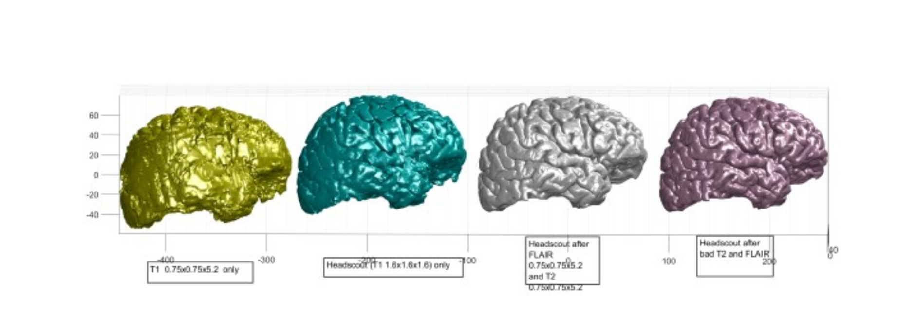
i) Start with the T1 headscout volume: 1.6mmx1.6mmx1.625mm This produces poor pial results but much better than using the T1s with a small number of slices. ii) Add in the flair data: Use -FLAIR and -autorecon3 with a FLAIR MRI scan This dramatically improves the pial surfaces and they look almost normal iii) Add some T2 data. This give more improvement of the pial surfaces
See attached image for how the cortex looks after these stages
My questions are:
*1) How can I add in the data from the higher resolution T1 scans? (with only 30 slices)*
*2) If I add a second T2 scan with -T2 -T2pial -autorecon3 will the results improve even more?*
Regards, Michael

External Email - Use Caution
I think there are some deep learning approaches out there that try to infer a high-resolution volume from low resolution volumes in multiple planes. I don’t know if they would work with only axial and sagittal images though.
Matt.
From: freesurfer-bounces@nmr.mgh.harvard.edu on behalf of Michael Mc Laughlin michael@thecurate.com Reply-To: Freesurfer support list Freesurfer@nmr.mgh.harvard.edu Date: Friday, March 31, 2023 at 6:36 AM To: Freesurfer support list Freesurfer@nmr.mgh.harvard.edu Subject: [Freesurfer] Is it possible to add additional T1 scans to improve surfaces/segmentation
External Email - Use Caution I am working with standard neuro radiological MRI scans.
These typically have something like this::
a) T1 headscout volume: 1.6mmx1.6mmx1.625mm 160x160x128 b) T1 se tra 0.72x0.72x5.2 320x320x30 c) T2 tse tra scan 0.72x0.72x5.2 320x320x30 d) T2 FLAIR (dark fluid) scan 0.58x0.58x5.2 460x460x30 c) T1 tse sag 0.72x0.72x5.2 320x320x30 c) T2 tse sag 0.72x0.72x5.2 320x320x30
I have found that the best results are obtained if I do the following:
i) Start with the T1 headscout volume: 1.6mmx1.6mmx1.625mm This produces poor pial results but much better than using the T1s with a small number of slices. ii) Add in the flair data: Use -FLAIR and -autorecon3 with a FLAIR MRI scan This dramatically improves the pial surfaces and they look almost normal iii) Add some T2 data. This give more improvement of the pial surfaces
See attached image for how the cortex looks after these stages
My questions are:
1) How can I add in the data from the higher resolution T1 scans? (with only 30 slices)
2) If I add a second T2 scan with -T2 -T2pial -autorecon3 will the results improve even more?
Regards, Michael
________________________________ The materials in this message are private and may contain Protected Healthcare Information or other information of a sensitive nature. If you are not the intended recipient, be advised that any unauthorized use, disclosure, copying or the taking of any action in reliance on the contents of this information is strictly prohibited. If you have received this email in error, please immediately notify the sender via telephone or return mail.
External Email - Use Caution
Freesurfer seem to be able to do it very well when I add the FLAIR and T2 scans. These have better axial resolution (up to 570x570) but there are only 30 slices yet the results dramatically improve,
1) Can I, with freesurfer, add a second, different, T2 scan (again high resolution in 2D but only 30 slices) 2) Can I, with freesurfer, add another T1 scan (also high resolution in 2D but only 30 slices)
Michael
On Fri, Mar 31, 2023 at 12:48 PM Glasser, Matt glasserm@wustl.edu wrote:
External Email - Use CautionI think there are some deep learning approaches out there that try to infer a high-resolution volume from low resolution volumes in multiple planes. I don’t know if they would work with only axial and sagittal images though.
Matt.
*From: *freesurfer-bounces@nmr.mgh.harvard.edu on behalf of Michael Mc Laughlin michael@thecurate.com *Reply-To: *Freesurfer support list Freesurfer@nmr.mgh.harvard.edu *Date: *Friday, March 31, 2023 at 6:36 AM *To: *Freesurfer support list Freesurfer@nmr.mgh.harvard.edu *Subject: *[Freesurfer] Is it possible to add additional T1 scans to improve surfaces/segmentation
External Email - Use Caution *I am working with standard neuro radiological MRI scans.
These typically have something like this::
a) T1 headscout volume: 1.6mmx1.6mmx1.625mm 160x160x128
b) T1 se tra 0.72x0.72x5.2 320x320x30
c) T2 tse tra scan 0.72x0.72x5.2 320x320x30
d) T2 FLAIR (dark fluid) scan 0.58x0.58x5.2 460x460x30
c) T1 tse sag 0.72x0.72x5.2 320x320x30
c) T2 tse sag 0.72x0.72x5.2 320x320x30
I have found that the best results are obtained if I do the following:
i) Start with the T1 headscout volume: 1.6mmx1.6mmx1.625mm
This produces poor pial results but much better than using the T1s with a small number of slices.
ii) Add in the flair data: Use -FLAIR and -autorecon3 with a FLAIR MRI scan
This dramatically improves the pial surfaces and they look almost normal
iii) Add some T2 data.
This give more improvement of the pial surfaces
See attached image for how the cortex looks after these stages
My questions are:
*1) How can I add in the data from the higher resolution T1 scans? (with only 30 slices)*
*2) If I add a second T2 scan with -T2 -T2pial -autorecon3 will the results improve even more?*
Regards,
Michael
The materials in this message are private and may contain Protected Healthcare Information or other information of a sensitive nature. If you are not the intended recipient, be advised that any unauthorized use, disclosure, copying or the taking of any action in reliance on the contents of this information is strictly prohibited. If you have received this email in error, please immediately notify the sender via telephone or return mail. _______________________________________________ Freesurfer mailing list Freesurfer@nmr.mgh.harvard.edu https://secure-web.cisco.com/1OMhg3PCPFqQ6pqYtWRpAZPucJ_AXPKNcKezpWv29mKcVHO... The information in this e-mail is intended only for the person to whom it is addressed. If you believe this e-mail was sent to you in error and the e-mail contains patient information, please contact the Mass General Brigham Compliance HelpLine at https://secure-web.cisco.com/1wZpTDsYG8zAoT0XahY5lDvErqYyHXhyGoNLhn0mjfJf6sv... < https://secure-web.cisco.com/1wZpTDsYG8zAoT0XahY5lDvErqYyHXhyGoNLhn0mjfJf6sv... . Please note that this e-mail is not secure (encrypted). If you do not wish to continue communication over unencrypted e-mail, please notify the sender of this message immediately. Continuing to send or respond to e-mail after receiving this message means you understand and accept this risk and wish to continue to communicate over unencrypted e-mail.

That is what mri_synthsr does (which is part of the FS distribution)
Cheers Bruce
From: freesurfer-bounces@nmr.mgh.harvard.edu freesurfer-bounces@nmr.mgh.harvard.edu On Behalf Of Glasser, Matt Sent: Friday, March 31, 2023 7:48 AM To: Freesurfer support list Freesurfer@nmr.mgh.harvard.edu Subject: Re: [Freesurfer] Is it possible to add additional T1 scans to improve surfaces/segmentation
External Email - Use Caution I think there are some deep learning approaches out there that try to infer a high-resolution volume from low resolution volumes in multiple planes. I don’t know if they would work with only axial and sagittal images though.
Matt.
From: <freesurfer-bounces@nmr.mgh.harvard.edumailto:freesurfer-bounces@nmr.mgh.harvard.edu> on behalf of Michael Mc Laughlin <michael@thecurate.commailto:michael@thecurate.com> Reply-To: Freesurfer support list <Freesurfer@nmr.mgh.harvard.edumailto:Freesurfer@nmr.mgh.harvard.edu> Date: Friday, March 31, 2023 at 6:36 AM To: Freesurfer support list <Freesurfer@nmr.mgh.harvard.edumailto:Freesurfer@nmr.mgh.harvard.edu> Subject: [Freesurfer] Is it possible to add additional T1 scans to improve surfaces/segmentation
External Email - Use Caution I am working with standard neuro radiological MRI scans.
These typically have something like this::
a) T1 headscout volume: 1.6mmx1.6mmx1.625mm 160x160x128 b) T1 se tra 0.72x0.72x5.2 320x320x30 c) T2 tse tra scan 0.72x0.72x5.2 320x320x30 d) T2 FLAIR (dark fluid) scan 0.58x0.58x5.2 460x460x30 c) T1 tse sag 0.72x0.72x5.2 320x320x30 c) T2 tse sag 0.72x0.72x5.2 320x320x30
I have found that the best results are obtained if I do the following:
i) Start with the T1 headscout volume: 1.6mmx1.6mmx1.625mm This produces poor pial results but much better than using the T1s with a small number of slices. ii) Add in the flair data: Use -FLAIR and -autorecon3 with a FLAIR MRI scan This dramatically improves the pial surfaces and they look almost normal iii) Add some T2 data. This give more improvement of the pial surfaces
See attached image for how the cortex looks after these stages
My questions are:
1) How can I add in the data from the higher resolution T1 scans? (with only 30 slices)
2) If I add a second T2 scan with -T2 -T2pial -autorecon3 will the results improve even more?
Regards, Michael
________________________________ The materials in this message are private and may contain Protected Healthcare Information or other information of a sensitive nature. If you are not the intended recipient, be advised that any unauthorized use, disclosure, copying or the taking of any action in reliance on the contents of this information is strictly prohibited. If you have received this email in error, please immediately notify the sender via telephone or return mail.
External Email - Use Caution
Cool!. That looks like exactly what I need. Thanks a lot Bruce!
Michael
On Fri, Mar 31, 2023 at 3:09 PM Fischl, Bruce R.,PHD < BFISCHL@mgh.harvard.edu> wrote:
That is what mri_synthsr does (which is part of the FS distribution)
Cheers
Bruce
*From:* freesurfer-bounces@nmr.mgh.harvard.edu < freesurfer-bounces@nmr.mgh.harvard.edu> *On Behalf Of *Glasser, Matt *Sent:* Friday, March 31, 2023 7:48 AM *To:* Freesurfer support list Freesurfer@nmr.mgh.harvard.edu *Subject:* Re: [Freesurfer] Is it possible to add additional T1 scans to improve surfaces/segmentation
External Email - Use Caution *I think there are some deep learning approaches out there that try to infer a high-resolution volume from low resolution volumes in multiple planes. I don’t know if they would work with only axial and sagittal images though.
Matt.
*From: *freesurfer-bounces@nmr.mgh.harvard.edu on behalf of Michael Mc Laughlin michael@thecurate.com *Reply-To: *Freesurfer support list Freesurfer@nmr.mgh.harvard.edu *Date: *Friday, March 31, 2023 at 6:36 AM *To: *Freesurfer support list Freesurfer@nmr.mgh.harvard.edu *Subject: *[Freesurfer] Is it possible to add additional T1 scans to improve surfaces/segmentation
External Email - Use Caution *I am working with standard neuro radiological MRI scans.
These typically have something like this::
a) T1 headscout volume: 1.6mmx1.6mmx1.625mm 160x160x128
b) T1 se tra 0.72x0.72x5.2 320x320x30
c) T2 tse tra scan 0.72x0.72x5.2 320x320x30
d) T2 FLAIR (dark fluid) scan 0.58x0.58x5.2 460x460x30
c) T1 tse sag 0.72x0.72x5.2 320x320x30
c) T2 tse sag 0.72x0.72x5.2 320x320x30
I have found that the best results are obtained if I do the following:
i) Start with the T1 headscout volume: 1.6mmx1.6mmx1.625mm
This produces poor pial results but much better than using the T1s with a small number of slices.
ii) Add in the flair data: Use -FLAIR and -autorecon3 with a FLAIR MRI scan
This dramatically improves the pial surfaces and they look almost normal
iii) Add some T2 data.
This give more improvement of the pial surfaces
See attached image for how the cortex looks after these stages
My questions are:
*1) How can I add in the data from the higher resolution T1 scans? (with only 30 slices)*
*2) If I add a second T2 scan with -T2 -T2pial -autorecon3 will the results improve even more?*
Regards,
Michael
The materials in this message are private and may contain Protected Healthcare Information or other information of a sensitive nature. If you are not the intended recipient, be advised that any unauthorized use, disclosure, copying or the taking of any action in reliance on the contents of this information is strictly prohibited. If you have received this email in error, please immediately notify the sender via telephone or return mail. _______________________________________________ Freesurfer mailing list Freesurfer@nmr.mgh.harvard.edu https://secure-web.cisco.com/1gSxl_fbgVfMdhhzq-QohkVfFjclDP_6wpXWwKe5Alg2kgs... The information in this e-mail is intended only for the person to whom it is addressed. If you believe this e-mail was sent to you in error and the e-mail contains patient information, please contact the Mass General Brigham Compliance HelpLine at https://secure-web.cisco.com/1oekB7qOGVKwKoW4Ok47DWT2zoowZJNX4tMHivea7UL264e... < https://secure-web.cisco.com/1oekB7qOGVKwKoW4Ok47DWT2zoowZJNX4tMHivea7UL264e... . Please note that this e-mail is not secure (encrypted). If you do not wish to continue communication over unencrypted e-mail, please notify the sender of this message immediately. Continuing to send or respond to e-mail after receiving this message means you understand and accept this risk and wish to continue to communicate over unencrypted e-mail.
freesurfer@nmr.mgh.harvard.edu
-
 Fischl, Bruce R.,PHD
 -
 Glasser, Matt
 -
 Michael Mc Laughlin