
External Email - Use Caution
I am working with standard neuro radiological MRI scans.
These typically have something like this::
a) T1 headscout volume: 1.6mmx1.6mmx1.625mm 160x160x128 b) T1 se tra 0.72x0.72x5.2 320x320x30 c) T2 tse tra scan 0.72x0.72x5.2 320x320x30 d) T2 FLAIR (dark fluid) scan 0.58x0.58x5.2 460x460x30 c) T1 tse sag 0.72x0.72x5.2 320x320x30 c) T2 tse sag 0.72x0.72x5.2 320x320x30
I have found that the best results are obtained if I do the following:
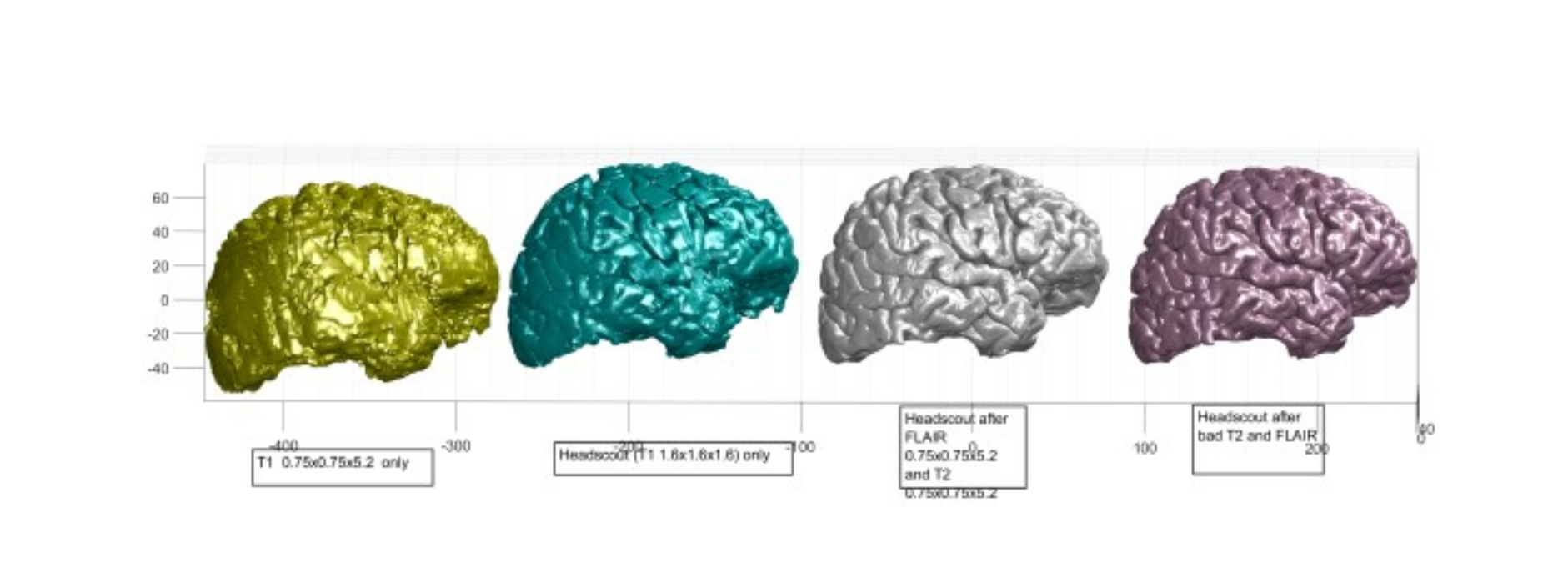
i) Start with the T1 headscout volume: 1.6mmx1.6mmx1.625mm This produces poor pial results but much better than using the T1s with a small number of slices. ii) Add in the flair data: Use -FLAIR and -autorecon3 with a FLAIR MRI scan This dramatically improves the pial surfaces and they look almost normal iii) Add some T2 data. This give more improvement of the pial surfaces
See attached image for how the cortex looks after these stages
My questions are:
*1) How can I add in the data from the higher resolution T1 scans? (with only 30 slices)*
*2) If I add a second T2 scan with -T2 -T2pial -autorecon3 will the results improve even more?*
Regards, Michael