
External Email - Use Caution
Dear all,
I am new to freesurfer and this might be an evident question, if so, I am sorry.
We have a longitudinal study with t0 and t1 scans. I followed the procedure and did the 3 steps.
1. Cross (with the following command: recon-all -subject 01_t0 -i /subjectfiles/t0/sanlm_13.nii -all -qcache -notal-check)
2. Base (recon-all -base 01_t0t1template -tp 01_t0 -tp 01_t1 -all)
3. Long (recon-all -long 01_t0 01_t0t1template -all)
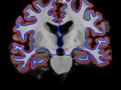
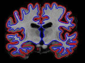
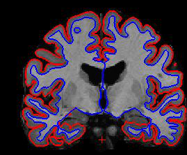
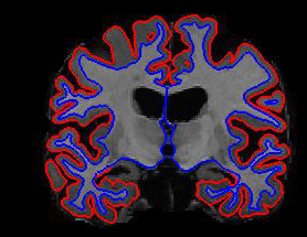
The problem is once I am inspecting the data, I see that the hippocampus is left out in all the cases (4 different subjects are attached in the screen captures).I used this command: freeview -v 01_t0/mri/norm.mgz \ -f 01_t0/surf/lh.pial:edgecolor=red \ 01_t0/surf/rh.pial:edgecolor=red \ 01_t0/surf/lh.white:edgecolor=blue \ 01_t0/surf/rh.white:edgecolor=blue)
Also, you should know that we used for T1 MP2RAGE instead of MPRAGE.
Is there anything I can do without having to re-do all the corrections by hand point by point and without having to restart over all my data pre-processing?
I have to specify that we are interested in the hippocampus, so this is a major point in our study.
Thanks in advance for your help,
Alba
[image: Screen Shot 2020-12-09 at 15.45.30.png] [image: Screen Shot 2020-12-09 at 15.45.38.png] [image: Screen Shot 2020-12-09 at 15.45.45.png] [image: Screen Shot 2020-12-09 at 15.45.55.png]

what do you mean the hippocampus is left out? it is not part of the surface since it is a subcortical structure. Look at the aseg.mgz
On 12/9/2020 9:53 AM, Albulena Shaqiri wrote:
External Email - Use Caution
Dear all,
I am new to freesurfer and this might be an evident question, if so, I am sorry.
We have a longitudinal study with t0 and t1 scans. I followed the procedure and did the 3 steps.
1. Cross (with the following command: recon-all -subject 01_t0 -i /subjectfiles/t0/sanlm_13.nii -all -qcache -notal-check)
2. Base (recon-all -base 01_t0t1template -tp 01_t0 -tp 01_t1 -all)
3. Long (recon-all -long 01_t0 01_t0t1template -all)
The problem is once I am inspecting the data, I see that the hippocampus is left out in all the cases (4 different subjects are attached in the screen captures).I used this command: freeview -v 01_t0/mri/norm.mgz \ -f 01_t0/surf/lh.pial:edgecolor=red \ 01_t0/surf/rh.pial:edgecolor=red \ 01_t0/surf/lh.white:edgecolor=blue \ 01_t0/surf/rh.white:edgecolor=blue)
Also, you should know that we used for T1 MP2RAGE instead of MPRAGE.
Is there anything I can do without having to re-do all the corrections by hand point by point and without having to restart over all my data pre-processing?
I have to specify that we are interested in the hippocampus, so this is a major point in our study.
Thanks in advance for your help,
Alba
[Screen Shot 2020-12-09 at 15.45.30.png] [Screen Shot 2020-12-09 at 15.45.38.png] [Screen Shot 2020-12-09 at 15.45.45.png] [Screen Shot 2020-12-09 at 15.45.55.png]
_______________________________________________ Freesurfer mailing list Freesurfer@nmr.mgh.harvard.edumailto:Freesurfer@nmr.mgh.harvard.edu https://mail.nmr.mgh.harvard.edu/mailman/listinfo/freesurfer
freesurfer@nmr.mgh.harvard.edu
-
 Albulena Shaqiri
 -
 Greve, Douglas N.,Ph.D.