
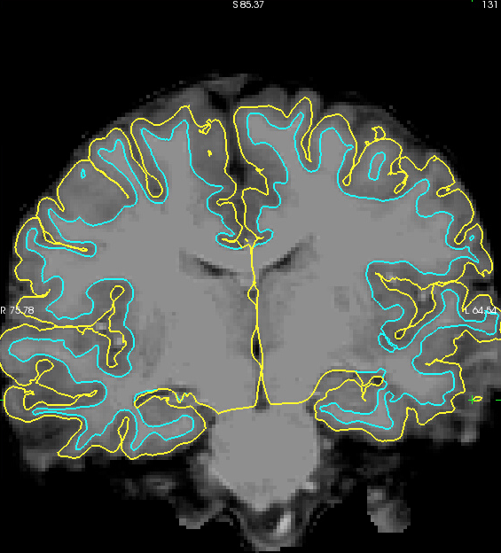
We have a few scans from kids that look like the image shown in the attached pic. We see a falloff in signal intensity in the gray matter that results in lots of cortex being excluded from the pial surface (e.g., along the dorsal surface and lateral temporal areas in the image). Typically, we would toss out this data and try to require from the subject or drop the subject from our analyses. But because of the age and clinical diagnosis of this subject we'd like to try to salvage this scan.
Can you offer any suggestions for how to improve the reconstruction? Is there a parameter that we can change that will allow the pial surface to fall farther from the white matter surface in terms of voxel intensity and distance? We would trade the need for more manual edits to get greater inclusion of cortex within the pial surface.
Do you have any tools or suggestions for a better intensity normalization of the original T1?
Thanks for any help that you can provide.
Best, Jason

Hello Jason,
The freesurfer folks may have some suggestions for adjusting certain parameters, but you can also try some denoising approaches before going into the freesurfer pipeline. There are a number of flavors. The link below is a good place to start I think.
https://www.sciencedirect.com/science/article/pii/S1361841515000171
Anthony -- Anthony Steven Dick, Ph.D. Associate Professor Director, Cognitive Neuroscience Program and Graduate Certificate in Cognitive Neuroscience Florida International University MM Campus AHC4 454 Department of Psychology and Center for Children and Familieshttp://www.ccf.fiu.edu/, an FIU Preeminent Programhttps://beyondpossible.fiu.edu/preeminent-programs/ 11200 S.W. 8th Street Miami, FL 33199 Ph: 305-348-4202; Lab Ph: 305-348-9055; Fx: 305-348-3879 Email: adick@fiu.edumailto:adick@fiu.edu; Webpage: http://dcn.fiu.eduhttp://dcn.fiu.edu/; Join the SSHDhttp://www.sshdonline.org/ ##### Check out the new bookhttp://www.routledge.com/9781138960039: Dick, A.S., & Müller, U. (2018). Advancing Developmental Science: Philosophy, Theory, and Method. Taylor & Francis.
From: freesurfer-bounces@nmr.mgh.harvard.edu on behalf of Jason Tourville jaytour@gmail.com Reply-To: Freesurfer support list freesurfer@nmr.mgh.harvard.edu Date: Thursday, February 1, 2018 at 11:43 AM To: Freesurfer support list freesurfer@nmr.mgh.harvard.edu Cc: Barbara Holland bobbiegholland@gmail.com Subject: [Freesurfer] options for a poor T1
We have a few scans from kids that look like the image shown in the attached pic. We see a falloff in signal intensity in the gray matter that results in lots of cortex being excluded from the pial surface (e.g., along the dorsal surface and lateral temporal areas in the image). Typically, we would toss out this data and try to require from the subject or drop the subject from our analyses. But because of the age and clinical diagnosis of this subject we'd like to try to salvage this scan.
Can you offer any suggestions for how to improve the reconstruction? Is there a parameter that we can change that will allow the pial surface to fall farther from the white matter surface in terms of voxel intensity and distance? We would trade the need for more manual edits to get greater inclusion of cortex within the pial surface.
Do you have any tools or suggestions for a better intensity normalization of the original T1?
Thanks for any help that you can provide.
Best, Jason
-- Jason A. Tourville, Ph.D. Research Assistant Professor Department of Speech, Language, and Hearing Sciences Boston University 677 Beacon St. Boston, MA 02215 Phone: (617)353-9484 Fax: (617)353-7755
freesurfer@nmr.mgh.harvard.edu
-
 Anthony Dick
 -
 Jason Tourville