
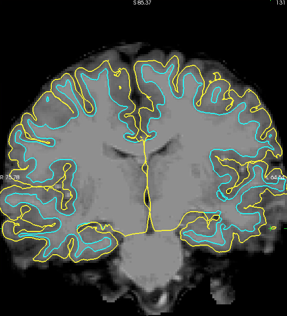
We have a few scans from kids that look like the image shown in the attached pic. We see a falloff in signal intensity in the gray matter that results in lots of cortex being excluded from the pial surface (e.g., along the dorsal surface and lateral temporal areas in the image). Typically, we would toss out this data and try to require from the subject or drop the subject from our analyses. But because of the age and clinical diagnosis of this subject we'd like to try to salvage this scan.
Can you offer any suggestions for how to improve the reconstruction? Is there a parameter that we can change that will allow the pial surface to fall farther from the white matter surface in terms of voxel intensity and distance? We would trade the need for more manual edits to get greater inclusion of cortex within the pial surface.
Do you have any tools or suggestions for a better intensity normalization of the original T1?
Thanks for any help that you can provide.
Best, Jason