
External Email - Use Caution
Dear Freesurfer Developers,
I have a question regarding volume analysis of fMRI data. I wanted to perform volume analysis in MNI305 space, and I used following command after creating a seed;
$mkanalysis-sess -analysis lh.pcc.mni305.vol -mni305 2 -stc siemens -fwhm 0 -notask -taskreg lh.pcc.dat 1 -nuisreg vcsf.dat 5 -nuisreg wm.dat 5 -mcextreg -polyfit 5 -nskip 10 -fsd rest -hpf 0.01 -TR 3 -per-run
$selxavg3-sess -s sess01 -a lh.pcc.mni305.vol -no-preproc -overwrite
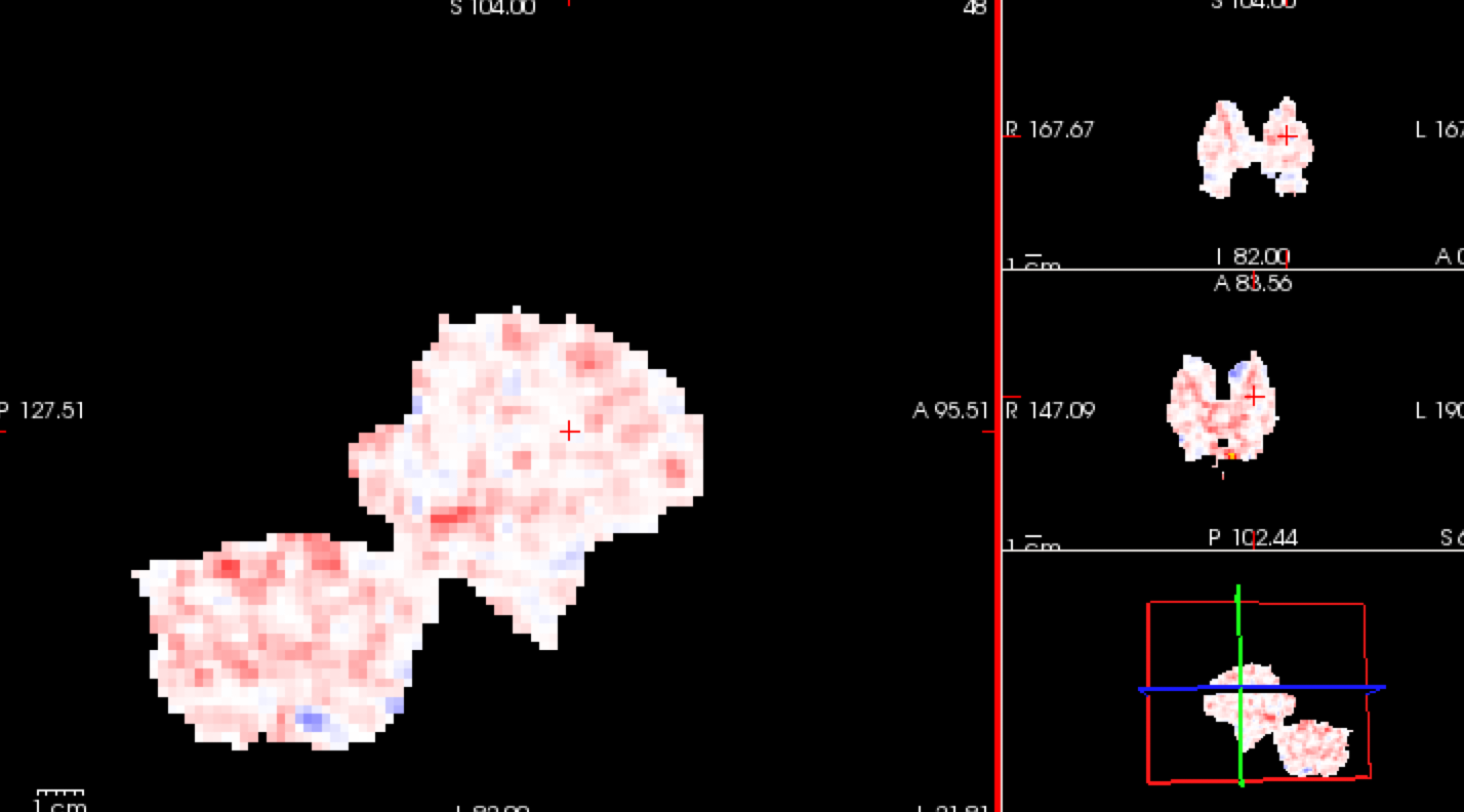
The resulting analysis seems only showing subcortical regions, not the whole brain. (Shown in the attachment) I read on the page that volume analysis is only for subcortical regions, is this the reason that I only see subcortical regions?
Another problem is that in the preprocessing step, I could not smooth the volume. If I do, it seems to distort the image and only gives something similar to the picture shown. Could this be the reason for the distorted analysis?
Thanks for your help.
Best, Wenzhen

Correct, we analyze the cortical surface using surface-based analysis. If you want to have full volume analysis, you can add -no-subcort-mask to preproc-sess. Not sure what the problem is with smoothing.
________________________________ From: freesurfer-bounces@nmr.mgh.harvard.edu freesurfer-bounces@nmr.mgh.harvard.edu on behalf of Wenzhen Zhao wenzhen.zhao@yale.edu Sent: Monday, October 5, 2020 3:22 PM To: Freesurfer support list freesurfer@nmr.mgh.harvard.edu Subject: [Freesurfer] Resting state fMRI - volume analysis
External Email - Use Caution
Dear Freesurfer Developers,
I have a question regarding volume analysis of fMRI data. I wanted to perform volume analysis in MNI305 space, and I used following command after creating a seed;
$mkanalysis-sess -analysis lh.pcc.mni305.vol -mni305 2 -stc siemens -fwhm 0 -notask -taskreg lh.pcc.dat 1 -nuisreg vcsf.dat 5 -nuisreg wm.dat 5 -mcextreg -polyfit 5 -nskip 10 -fsd rest -hpf 0.01 -TR 3 -per-run
$selxavg3-sess -s sess01 -a lh.pcc.mni305.vol -no-preproc -overwrite
The resulting analysis seems only showing subcortical regions, not the whole brain. (Shown in the attachment) I read on the page that volume analysis is only for subcortical regions, is this the reason that I only see subcortical regions?
Another problem is that in the preprocessing step, I could not smooth the volume. If I do, it seems to distort the image and only gives something similar to the picture shown. Could this be the reason for the distorted analysis?
Thanks for your help.
Best, Wenzhen
[cid:1B7549AE-41DD-4BB5-AFE3-BFC8DE37DEEA@its.yale.edu]
External Email - Use Caution
Thank you for your help.
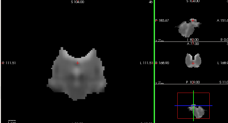
The smoothing issue occurs only on volume. It works okay with surface. When I smooth the volume by 5, the results look like below. The command I used was $preproc-sess -s sess01 -fsd rest -stc siemens -noreg -mni305 -vol-fwhm 5 -no-subcort-mask -per-run -update
Thanks, Wenzhen
On Oct 6, 2020, at 11:53 AM, Greve, Douglas N.,Ph.D. DGREVE@mgh.harvard.edu wrote:
Correct, we analyze the cortical surface using surface-based analysis. If you want to have full volume analysis, you can add -no-subcort-mask to preproc-sess. Not sure what the problem is with smoothing.
From: freesurfer-bounces@nmr.mgh.harvard.edu mailto:freesurfer-bounces@nmr.mgh.harvard.edu <freesurfer-bounces@nmr.mgh.harvard.edu mailto:freesurfer-bounces@nmr.mgh.harvard.edu> on behalf of Wenzhen Zhao <wenzhen.zhao@yale.edu mailto:wenzhen.zhao@yale.edu> Sent: Monday, October 5, 2020 3:22 PM To: Freesurfer support list <freesurfer@nmr.mgh.harvard.edu mailto:freesurfer@nmr.mgh.harvard.edu> Subject: [Freesurfer] Resting state fMRI - volume analysis
External Email - Use CautionDear Freesurfer Developers,
I have a question regarding volume analysis of fMRI data. I wanted to perform volume analysis in MNI305 space, and I used following command after creating a seed;
$mkanalysis-sess -analysis lh.pcc.mni305.vol -mni305 2 -stc siemens -fwhm 0 -notask -taskreg lh.pcc.dat 1 -nuisreg vcsf.dat 5 -nuisreg wm.dat 5 -mcextreg -polyfit 5 -nskip 10 -fsd rest -hpf 0.01 -TR 3 -per-run
$selxavg3-sess -s sess01 -a lh.pcc.mni305.vol -no-preproc -overwrite
The resulting analysis seems only showing subcortical regions, not the whole brain. (Shown in the attachment) I read on the page that volume analysis is only for subcortical regions, is this the reason that I only see subcortical regions?
Another problem is that in the preprocessing step, I could not smooth the volume. If I do, it seems to distort the image and only gives something similar to the picture shown. Could this be the reason for the distorted analysis?
Thanks for your help.
Best, Wenzhen
<Screen Shot 2020-10-05 at 2.31.29 PM.png> _______________________________________________ Freesurfer mailing list Freesurfer@nmr.mgh.harvard.edu mailto:Freesurfer@nmr.mgh.harvard.edu https://mail.nmr.mgh.harvard.edu/mailman/listinfo/freesurfer https://mail.nmr.mgh.harvard.edu/mailman/listinfo/freesurfer
What is wrong?


On 10/6/2020 12:28 PM, Wenzhen Zhao wrote:
        External Email - Use Caution

Thank you for your help.

The smoothing issue occurs only on volume. It works okay with
surface. When I smooth the volume by 5, the results look like
below. The command I used was $preproc-sess -s sess01 -fsd rest
-stc siemens -noreg  -mni305 -vol-fwhm 5 -no-subcort-mask
-per-run -update


Thanks,
Wenzhen
On Oct 6, 2020, at 11:53 AM, Greve, Douglas N.,Ph.D. <DGREVE@mgh.harvard.edu mailto:DGREVE@mgh.harvard.edu> wrote:
Correct, we analyze the cortical surface using surface-based analysis. If you want to have full volume analysis, you can add -no-subcort-mask to preproc-sess. Not sure what the problem is with smoothing.
*From:*freesurfer-bounces@nmr.mgh.harvard.edu mailto:freesurfer-bounces@nmr.mgh.harvard.edu<freesurfer-bounces@nmr.mgh.harvard.edu mailto:freesurfer-bounces@nmr.mgh.harvard.edu> on behalf of Wenzhen Zhao <wenzhen.zhao@yale.edu mailto:wenzhen.zhao@yale.edu> *Sent:*Monday, October 5, 2020 3:22 PM *To:*Freesurfer support list <freesurfer@nmr.mgh.harvard.edu mailto:freesurfer@nmr.mgh.harvard.edu> *Subject:*[Freesurfer] Resting state fMRI - volume analysis External Email - Use Caution Dear Freesurfer Developers,
I have a question regarding volume analysis of fMRI data. I wanted to perform volume analysis in MNI305 space, and I used following command after creating a seed;
$mkanalysis-sess -analysis lh.pcc.mni305.vol -mni305 2 -stc siemens -fwhm 0 -notask -taskreg lh.pcc.dat 1 -nuisreg vcsf.dat 5 -nuisreg wm.dat 5 -mcextreg -polyfit 5 -nskip 10 -fsd rest -hpf 0.01 -TR 3 -per-run
$selxavg3-sess -s sess01 -a lh.pcc.mni305.vol -no-preproc -overwrite
The resulting analysis seems only showing subcortical regions, not the whole brain. (Shown in the attachment) I read on the page that volume analysis is only for subcortical regions, is this the reason that I only see subcortical regions?
Another problem is that in the preprocessing step, I could not smooth the volume. If I do, it seems to distort the image and only gives something similar to the picture shown. Could this be the reason for the distorted analysis?
Thanks for your help.
Best, Wenzhen
<Screen Shot 2020-10-05 at 2.31.29 PM.png> _______________________________________________ Freesurfer mailing list Freesurfer@nmr.mgh.harvard.edu mailto:Freesurfer@nmr.mgh.harvard.edu https://mail.nmr.mgh.harvard.edu/mailman/listinfo/freesurfer
Freesurfer mailing list Freesurfer@nmr.mgh.harvard.edu https://mail.nmr.mgh.harvard.edu/mailman/listinfo/freesurfer
External Email - Use Caution
Hi,
I find it weird that it does not show full brain after smoothing (-fwhm 5). Is this normal?
Also, even with -no-subcort-mask, volume analysis does not do full brain analysis.
Best, Wenzhen
On Wed, Oct 7, 2020 at 10:16 AM Douglas N. Greve dgreve@mgh.harvard.edu wrote:
What is wrong?
On 10/6/2020 12:28 PM, Wenzhen Zhao wrote:
External Email - Use CautionThank you for your help.
The smoothing issue occurs only on volume. It works okay with surface. When I smooth the volume by 5, the results look like below. The command I used was $preproc-sess -s sess01 -fsd rest -stc siemens -noreg -mni305 -vol-fwhm 5 -no-subcort-mask -per-run -update
Thanks, Wenzhen
On Oct 6, 2020, at 11:53 AM, Greve, Douglas N.,Ph.D. < DGREVE@mgh.harvard.edu> wrote:
Correct, we analyze the cortical surface using surface-based analysis. If you want to have full volume analysis, you can add -no-subcort-mask to preproc-sess. Not sure what the problem is with smoothing.
*From:* freesurfer-bounces@nmr.mgh.harvard.edu < freesurfer-bounces@nmr.mgh.harvard.edu> on behalf of Wenzhen Zhao < wenzhen.zhao@yale.edu> *Sent:* Monday, October 5, 2020 3:22 PM *To:* Freesurfer support list freesurfer@nmr.mgh.harvard.edu *Subject:* [Freesurfer] Resting state fMRI - volume analysis
External Email - Use CautionDear Freesurfer Developers,
I have a question regarding volume analysis of fMRI data. I wanted to perform volume analysis in MNI305 space, and I used following command after creating a seed;
$mkanalysis-sess -analysis lh.pcc.mni305.vol -mni305 2 -stc siemens -fwhm 0 -notask -taskreg lh.pcc.dat 1 -nuisreg vcsf.dat 5 -nuisreg wm.dat 5 -mcextreg -polyfit 5 -nskip 10 -fsd rest -hpf 0.01 -TR 3 -per-run
$selxavg3-sess -s sess01 -a lh.pcc.mni305.vol -no-preproc -overwrite
The resulting analysis seems only showing subcortical regions, not the whole brain. (Shown in the attachment) I read on the page that volume analysis is only for subcortical regions, is this the reason that I only see subcortical regions?
Another problem is that in the preprocessing step, I could not smooth the volume. If I do, it seems to distort the image and only gives something similar to the picture shown. Could this be the reason for the distorted analysis?
Thanks for your help.
Best, Wenzhen
<Screen Shot 2020-10-05 at 2.31.29 PM.png> _______________________________________________ Freesurfer mailing list Freesurfer@nmr.mgh.harvard.edu https://mail.nmr.mgh.harvard.edu/mailman/listinfo/freesurfer
Freesurfer mailing listFreesurfer@nmr.mgh.harvard.eduhttps://mail.nmr.mgh.harvard.edu/mailman/listinfo/freesurfer
Freesurfer mailing list Freesurfer@nmr.mgh.harvard.edu https://mail.nmr.mgh.harvard.edu/mailman/listinfo/freesurfer
Did you see the part about only analyzing subcortical gray matter earlier in our conversation?
On 10/7/2020 12:00 PM, Wenzhen Zhao wrote:
External Email - Use Caution
Hi,
I find it weird that it does not show full brain after smoothing (-fwhm 5). Is this normal?
Also, even with -no-subcort-mask, volume analysis does not do full brain analysis.
Best, Wenzhen
On Wed, Oct 7, 2020 at 10:16 AM Douglas N. Greve <dgreve@mgh.harvard.edu mailto:dgreve@mgh.harvard.edu> wrote:
What is wrong? On 10/6/2020 12:28 PM, Wenzhen Zhao wrote:External Email - Use Caution Thank you for your help. The smoothing issue occurs only on volume. It works okay with surface. When I smooth the volume by 5, the results look like below. The command I used was $preproc-sess -s sess01 -fsd rest -stc siemens -noreg -mni305 -vol-fwhm 5 -no-subcort-mask -per-run -update Thanks, WenzhenOn Oct 6, 2020, at 11:53 AM, Greve, Douglas N.,Ph.D. <DGREVE@mgh.harvard.edu <mailto:DGREVE@mgh.harvard.edu>> wrote: Correct, we analyze the cortical surface using surface-based analysis. If you want to have full volume analysis, you can add -no-subcort-mask to preproc-sess. Not sure what the problem is with smoothing.------------------------------------------------------------------------ *From:*freesurfer-bounces@nmr.mgh.harvard.edu <mailto:freesurfer-bounces@nmr.mgh.harvard.edu><freesurfer-bounces@nmr.mgh.harvard.edu <mailto:freesurfer-bounces@nmr.mgh.harvard.edu>> on behalf of Wenzhen Zhao <wenzhen.zhao@yale.edu <mailto:wenzhen.zhao@yale.edu>> *Sent:*Monday, October 5, 2020 3:22 PM *To:*Freesurfer support list <freesurfer@nmr.mgh.harvard.edu <mailto:freesurfer@nmr.mgh.harvard.edu>> *Subject:*[Freesurfer] Resting state fMRI - volume analysis External Email - Use Caution Dear Freesurfer Developers, I have a question regarding volume analysis of fMRI data. I wanted to perform volume analysis in MNI305 space, and I used following command after creating a seed; $mkanalysis-sess -analysis lh.pcc.mni305.vol -mni305 2 -stc siemens -fwhm 0 -notask -taskreg lh.pcc.dat 1 -nuisreg vcsf.dat 5 -nuisreg wm.dat 5 -mcextreg -polyfit 5 -nskip 10 -fsd rest -hpf 0.01 -TR 3 -per-run $selxavg3-sess -s sess01 -a lh.pcc.mni305.vol -no-preproc -overwrite The resulting analysis seems only showing subcortical regions, not the whole brain. (Shown in the attachment) I read on the page that volume analysis is only for subcortical regions, is this the reason that I only see subcortical regions? Another problem is that in the preprocessing step, I could not smooth the volume. If I do, it seems to distort the image and only gives something similar to the picture shown. Could this be the reason for the distorted analysis? Thanks for your help. Best, Wenzhen <Screen Shot 2020-10-05 at 2.31.29 PM.png> _______________________________________________ Freesurfer mailing list Freesurfer@nmr.mgh.harvard.edu <mailto:Freesurfer@nmr.mgh.harvard.edu> https://mail.nmr.mgh.harvard.edu/mailman/listinfo/freesurfer_______________________________________________ Freesurfer mailing list Freesurfer@nmr.mgh.harvard.edu <mailto:Freesurfer@nmr.mgh.harvard.edu> https://mail.nmr.mgh.harvard.edu/mailman/listinfo/freesurfer_______________________________________________ Freesurfer mailing list Freesurfer@nmr.mgh.harvard.edu <mailto:Freesurfer@nmr.mgh.harvard.edu> https://mail.nmr.mgh.harvard.edu/mailman/listinfo/freesurfer
Freesurfer mailing list Freesurfer@nmr.mgh.harvard.edu https://mail.nmr.mgh.harvard.edu/mailman/listinfo/freesurfer
freesurfer@nmr.mgh.harvard.edu
-
 Douglas N. Greve
 -
Greve, Douglas N.,Ph.D.
-
 Wenzhen Zhao