
Correct, we analyze the cortical surface using surface-based analysis. If you want to have full volume analysis, you can add -no-subcort-mask to preproc-sess. Not sure what the problem is with smoothing.
________________________________ From: freesurfer-bounces@nmr.mgh.harvard.edu freesurfer-bounces@nmr.mgh.harvard.edu on behalf of Wenzhen Zhao wenzhen.zhao@yale.edu Sent: Monday, October 5, 2020 3:22 PM To: Freesurfer support list freesurfer@nmr.mgh.harvard.edu Subject: [Freesurfer] Resting state fMRI - volume analysis
External Email - Use Caution
Dear Freesurfer Developers,
I have a question regarding volume analysis of fMRI data. I wanted to perform volume analysis in MNI305 space, and I used following command after creating a seed;
$mkanalysis-sess -analysis lh.pcc.mni305.vol -mni305 2 -stc siemens -fwhm 0 -notask -taskreg lh.pcc.dat 1 -nuisreg vcsf.dat 5 -nuisreg wm.dat 5 -mcextreg -polyfit 5 -nskip 10 -fsd rest -hpf 0.01 -TR 3 -per-run
$selxavg3-sess -s sess01 -a lh.pcc.mni305.vol -no-preproc -overwrite
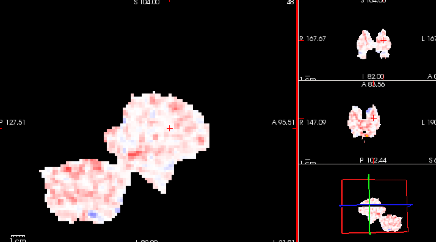
The resulting analysis seems only showing subcortical regions, not the whole brain. (Shown in the attachment) I read on the page that volume analysis is only for subcortical regions, is this the reason that I only see subcortical regions?
Another problem is that in the preprocessing step, I could not smooth the volume. If I do, it seems to distort the image and only gives something similar to the picture shown. Could this be the reason for the distorted analysis?
Thanks for your help.
Best, Wenzhen
[cid:1B7549AE-41DD-4BB5-AFE3-BFC8DE37DEEA@its.yale.edu]