
Hi Mike
I'm not sure - I think the insula was an additional label that Ron Killiany added. I'll cc Ron to see if he has any insight. It certainly looks too medial to me.
I would also check on the white surface just to make sure that it's not a quirk of the inflation. If the aseg looks right we could certainly put together some simple auto-editing to correct this kind of thing
cheers Bruce
On Thu, 7 Aug 2014, Harms, Michael wrote:
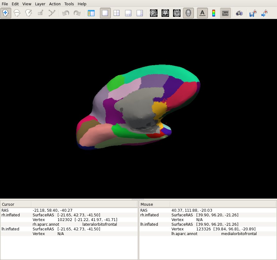
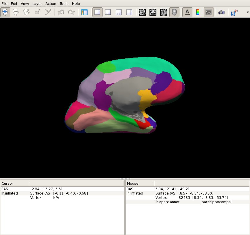
Hi, An astute RA, while reviewing the aparc+aseg.mgz of brains processed with FS 5.3, noticed a fair number of instances, where the insula label "wraps around" pretty medially, where she was expecting to see orbitofrontal as the label. See attached jpg's for two examples shown on the inflated surface. Since this is fairly common, I'm assuming that this isn't anything to be concerned about, but just wanted to confirm.
Has this always been the behavior of the insula label, and we just didn't notice it previously?
thanks, -MH
-- Michael Harms, Ph.D.
Conte Center for the Neuroscience of Mental Disorders Washington University School of Medicine Department of Psychiatry, Box 8134 660 South Euclid Ave. Tel: 314-747-6173 St. Louis, MO 63110 Email: mharms@wustl.edu
The materials in this message are private and may contain Protected Healthcare Information or other information of a sensitive nature. If you are not the intended recipient, be advised that any unauthorized use, disclosure, copying or the taking of any action in reliance on the contents of this information is strictly prohibited. If you have received this email in error, please immediately notify the sender via telephone or return mail.

I asked a neuroanatomist and insula expert on my campus about this a while back and this was his response:
"This is the region of the "limen insulae" that you will remember Dr. Heimer referring to often in the brain dissection course. The insula is defined as cortex which is covered by a lid of other cortical lobes, frontal, parietal and temporal. They all fold over except at the inferior frontal end where a little bit of the surface of otherwise insular cortex is not covered, and this little corner peeks out. This is the limeninsula which neighbors the olfactory cortex on the inferior surface of the frontal lobe and the medial surface of the temporal lobe, and is olfactory cortex itself. Since it is not covered over, it technically might not be considered really "insular" but it is obviously in continuity with, and not separated by any kind of sulcus or other landmark, from the rest of the insular covered cortex. So we are left with no secure boundary line between insular cortex and inferior frontal and medial temporal cortex in this corner. It looks like Freesurfer has decided to call it all insular, and with good functional reasons."
Cheers,
On Thu, Aug 7, 2014 at 3:32 PM, Bruce Fischl fischl@nmr.mgh.harvard.edu wrote:
Hi Mike
I'm not sure - I think the insula was an additional label that Ron Killiany added. I'll cc Ron to see if he has any insight. It certainly looks too medial to me.
I would also check on the white surface just to make sure that it's not a quirk of the inflation. If the aseg looks right we could certainly put together some simple auto-editing to correct this kind of thing
cheers Bruce
On Thu, 7 Aug 2014, Harms, Michael wrote:
Hi, An astute RA, while reviewing the aparc+aseg.mgz of brains processed with FS 5.3, noticed a fair number of instances, where the insula label "wraps around" pretty medially, where she was expecting to see orbitofrontal as the label. See attached jpg's for two examples shown on the inflated surface. Since this is fairly common, I'm assuming that this isn't anything to be concerned about, but just wanted to confirm.
Has this always been the behavior of the insula label, and we just didn't notice it previously?
thanks, -MH
-- Michael Harms, Ph.D.
Conte Center for the Neuroscience of Mental Disorders Washington University School of Medicine Department of Psychiatry, Box 8134 660 South Euclid Ave. Tel: 314-747-6173 St. Louis, MO 63110 Email: mharms@wustl.edu
The materials in this message are private and may contain Protected Healthcare Information or other information of a sensitive nature. If you are not the intended recipient, be advised that any unauthorized use, disclosure, copying or the taking of any action in reliance on the contents of this information is strictly prohibited. If you have received this email in error, please immediately notify the sender via telephone or return mail.
Freesurfer mailing list Freesurfer@nmr.mgh.harvard.edu https://mail.nmr.mgh.harvard.edu/mailman/listinfo/freesurfer
The information in this e-mail is intended only for the person to whom it is addressed. If you believe this e-mail was sent to you in error and the e-mail contains patient information, please contact the Partners Compliance HelpLine at http://www.partners.org/complianceline . If the e-mail was sent to you in error but does not contain patient information, please contact the sender and properly dispose of the e-mail.
wow, I like that answer much more than "it's a bug"! Can we go with Paul's explanation?
Bruce
On Thu, 7 Aug 2014, Paul Beach wrote:
I asked a neuroanatomist and insula expert on my campus about this a while back and this was his response: "This is the region of the "limen insulae" that you will remember Dr. Heimer referring to often in the brain dissection course. The insula is defined as cortex which is covered by a lid of other cortical lobes, frontal, parietal and temporal. They all fold over except at the inferior frontal end where a little bit of the surface of otherwise insular cortex is not covered, and this little corner peeks out. This is the limeninsula which neighbors the olfactory cortex on the inferior surface of the frontal lobe and the medial surface of the temporal lobe, and is olfactory cortex itself. Since it is not covered over, it technically might not be considered really "insular" but it is obviously in continuity with, and not separated by any kind of sulcus or other landmark, from the rest of the insular covered cortex. So we are left with no secure boundary line between insular cortex and inferior frontal and medial temporal cortex in this corner. It looks like Freesurfer has decided to call it all insular, and with good functional reasons."
Cheers,
On Thu, Aug 7, 2014 at 3:32 PM, Bruce Fischl fischl@nmr.mgh.harvard.edu wrote: Hi Mike
  I'm not sure - I think the insula was an additional label that
  Ron Killiany added. I'll cc Ron to see if he has any insight. It
  certainly looks too medial to me.

  I would also check on the white surface just to make sure that
  it's not a quirk of the inflation. If the aseg looks right we
  could certainly put together some simple auto-editing to correct
  this kind of thing

  cheers
  Bruce


  On Thu, 7 Aug 2014, Harms, Michael wrote:


  Hi,
  An astute RA, while reviewing the aparc+aseg.mgz of brains
  processed with FS
  5.3, noticed a fair number of instances, where the insula
  label "wraps
  around" pretty medially, where she was expecting to see
  orbitofrontal as the
  label.  See attached jpg's for two examples shown on the
  inflated surface.
   Since this is fairly common, I'm assuming that this isn't
  anything to be
  concerned about, but just wanted to confirm.

  Has this always been the behavior of the insula label, and
  we just didn't
  notice it previously?

  thanks,
  -MH

  --
  Michael Harms, Ph.D.
  -----------------------------------------------------------
  Conte Center for the Neuroscience of Mental Disorders
  Washington University School of Medicine
  Department of Psychiatry, Box 8134
  660 South Euclid Ave. Tel: 314-747-6173
  St. Louis, MO  63110 Email: mharms@wustl.edu
_
The materials in this message are private and may contain Protected Healthcare Information or other information of a sensitive nature. If you are not the intended recipient, be advised that any unauthorized use, disclosure, copying or the taking of any action in reliance on the contents of this information is strictly prohibited. If you have received this email in error, please immediately notify the sender via telephone or return mail.
Freesurfer mailing list Freesurfer@nmr.mgh.harvard.edu https://mail.nmr.mgh.harvard.edu/mailman/listinfo/freesurfer
The information in this e-mail is intended only for the person to whom it is addressed. If you believe this e-mail was sent to you in error and the e-mail contains patient information, please contact the Partners Compliance HelpLine at http://www.partners.org/complianceline . If the e-mail was sent to you in error but does not contain patient information, please contact the sender and properly dispose of the e-mail.
-- Paul Beach DO/PhD candidate - Year VI Michigan State University
- College of Osteopathic Medicine
- Neuroscience Program - MSU Cognitive and Geriatric Neurology Team (CoGeNT)

I'm not itching to re-process this data so I like that explanation as well. I think my main concern is that that bit of insula label seems somewhat arbitrary and inconsistent, in that some subjects have this bit of medial tissue labelled as insula, whereas others don't. (Even within the same subject, it isn't consistent between the two hemispheres).
I guess it is just a matter of the sulcal variability and atlas labeling resulting in variability in that particular region. Presumably, Ron must have labelled that bit of tissue as "insula" in some subjects, but not others??
Ron: It would be great to hear what kind of "rule" you applied when labeling the individual subjects in the atlas in that particular region when deciding whether it should be considered insula or medial/lateral orbitofrontal.
thanks, -MH
-- Michael Harms, Ph.D.
----------------------------------------------------------- Conte Center for the Neuroscience of Mental Disorders Washington University School of Medicine Department of Psychiatry, Box 8134 660 South Euclid Ave. Tel: 314-747-6173 St. Louis, MO 63110 Email: mharms@wustl.edu
On 8/7/14 2:52 PM, "Bruce Fischl" fischl@nmr.mgh.harvard.edu wrote:
wow, I like that answer much more than "it's a bug"! Can we go with Paul's explanation?
Bruce
On Thu, 7 Aug 2014, Paul Beach wrote:
I asked a neuroanatomist and insula expert on my campus about this a while back and this was his response: "This is the region of the "limen insulae" that you will remember Dr. Heimer referring to often in the brain dissection course. The insula is defined as cortex which is covered by a lid of other cortical lobes, frontal, parietal and temporal. They all fold over except at the inferior frontal end where a little bit of the surface of otherwise insular cortex is not covered, and this little corner peeks out. This is the limeninsula which neighbors the olfactory cortex on the inferior surface of the frontal lobe and the medial surface of the temporal lobe, and is olfactory cortex itself. Since it is not covered over, it technically might not be considered really "insular" but it is obviously in continuity with, and not separated by any kind of sulcus or other landmark, from the rest of the insular covered cortex. So we are left with no secure boundary line between insular cortex and inferior frontal and medial temporal cortex in this corner. It looks like Freesurfer has decided to call it all insular, and with good functional reasons."
Cheers,
On Thu, Aug 7, 2014 at 3:32 PM, Bruce Fischl fischl@nmr.mgh.harvard.edu wrote: Hi Mike
  I'm not sure - I think the insula was an additional label that
  Ron Killiany added. I'll cc Ron to see if he has any insight. It
  certainly looks too medial to me.

  I would also check on the white surface just to make sure that
  it's not a quirk of the inflation. If the aseg looks right we
  could certainly put together some simple auto-editing to correct
  this kind of thing

  cheers
  Bruce


  On Thu, 7 Aug 2014, Harms, Michael wrote:


  Hi,
  An astute RA, while reviewing the aparc+aseg.mgz of brains
  processed with FS
  5.3, noticed a fair number of instances, where the insula
  label "wraps
  around" pretty medially, where she was expecting to see
  orbitofrontal as the
  label.  See attached jpg's for two examples shown on the
  inflated surface.
   Since this is fairly common, I'm assuming that this isn't
  anything to be
  concerned about, but just wanted to confirm.

  Has this always been the behavior of the insula label, and
  we just didn't
  notice it previously?

  thanks,
  -MH

  --
  Michael Harms, Ph.D.
  -----------------------------------------------------------
  Conte Center for the Neuroscience of Mental Disorders
  Washington University School of Medicine
  Department of Psychiatry, Box 8134
  660 South Euclid Ave. Tel: 314-747-6173
  St. Louis, MO  63110 Email: mharms@wustl.edu
__ _
The materials in this message are private and may contain Protected Healthcare Information or other information of a sensitive nature. If you are not the intended recipient, be advised that any unauthorized use, disclosure, copying or the taking of any action in reliance on the contents of this information is strictly prohibited. If you have received this email in error, please immediately notify the sender via telephone or return mail.
Freesurfer mailing list Freesurfer@nmr.mgh.harvard.edu https://mail.nmr.mgh.harvard.edu/mailman/listinfo/freesurfer
The information in this e-mail is intended only for the person to whom it is addressed. If you believe this e-mail was sent to you in error and the e-mail contains patient information, please contact the Partners Compliance HelpLine at http://www.partners.org/complianceline . If the e-mail was sent to you in error but does not contain patient information, please contact the sender and properly dispose of the e-mail.
-- Paul Beach DO/PhD candidate - Year VI Michigan State University
- College of Osteopathic Medicine
- Neuroscience Program - MSU Cognitive and Geriatric Neurology Team
(CoGeNT)
________________________________ The materials in this message are private and may contain Protected Healthcare Information or other information of a sensitive nature. If you are not the intended recipient, be advised that any unauthorized use, disclosure, copying or the taking of any action in reliance on the contents of this information is strictly prohibited. If you have received this email in error, please immediately notify the sender via telephone or return mail.

Hi Michael the limen insula explanation sounds ok, even if it's extension on the medial surface seem really large. would you have a snapshot to look on the volume to which structure this label anatomically corresponds to ? Also did you had a look to the results of the insula aparc with the 2009a atlas? cd
Hi Christophe, I uploaded both subjects that I showed the snapshots for yesterday into the FS FTP drop site. (Do you have access to that?)
cheers, -MH
-- Michael Harms, Ph.D.
----------------------------------------------------------- Conte Center for the Neuroscience of Mental Disorders Washington University School of Medicine Department of Psychiatry, Box 8134 660 South Euclid Ave. Tel: 314-747-6173 St. Louis, MO 63110 Email: mharms@wustl.edu
On 8/7/14 4:03 PM, "Christophe Destrieux" christophe.destrieux@univ-tours.fr wrote:
Hi Michael the limen insula explanation sounds ok, even if it's extension on the medial surface seem really large. would you have a snapshot to look on the volume to which structure this label anatomically corresponds to ? Also did you had a look to the results of the insula aparc with the 2009a atlas? cd
-- Christophe Destrieux,
Directeur du Département Communication et Multimédia http://med.univ-tours.fr/M0S01/0/fiche___defaultstructureksup/&RH=120065... 59612
Unité « Imagerie et Cerveau » UMRS INSERM U930, Université François Rabelais de Tours http://www.u930.tours.inserm.fr/
Service de Neurochirurgie et Laboratoire d'Anatomie - Faculté de Médecine - 10 Bd Tonnellé 37032 Tours - France tel (33) 2 47 36 61 36 - fax (33) 2 47 36 62 07
----- Mail original ----- De: "Michael Harms" mharms@wustl.edu À: "Freesurfer support list" freesurfer@nmr.mgh.harvard.edu Cc: "Ron KILLIANY" rkilliany@cajal-1.bu.edu Envoyé: Jeudi 7 Août 2014 22:18:49 Objet: Re: [Freesurfer] insula on medial surface
I'm not itching to re-process this data so I like that explanation as well. I think my main concern is that that bit of insula label seems somewhat arbitrary and inconsistent, in that some subjects have this bit of medial tissue labelled as insula, whereas others don't. (Even within the same subject, it isn't consistent between the two hemispheres).
I guess it is just a matter of the sulcal variability and atlas labeling resulting in variability in that particular region. Presumably, Ron must have labelled that bit of tissue as "insula" in some subjects, but not others??
Ron: It would be great to hear what kind of "rule" you applied when labeling the individual subjects in the atlas in that particular region when deciding whether it should be considered insula or medial/lateral orbitofrontal.
thanks, -MH
-- Michael Harms, Ph.D.
Conte Center for the Neuroscience of Mental Disorders Washington University School of Medicine Department of Psychiatry, Box 8134 660 South Euclid Ave. Tel: 314-747-6173 St. Louis, MO 63110 Email: mharms@wustl.edu
On 8/7/14 2:52 PM, "Bruce Fischl" fischl@nmr.mgh.harvard.edu wrote:
wow, I like that answer much more than "it's a bug"! Can we go with Paul's explanation?
Bruce
On Thu, 7 Aug 2014, Paul Beach wrote:
I asked a neuroanatomist and insula expert on my campus about this a while back and this was his response: "This is the region of the "limen insulae" that you will remember Dr. Heimer referring to often in the brain dissection course. The insula is defined as cortex which is covered by a lid of other cortical lobes, frontal, parietal and temporal. They all fold over except at the inferior frontal end where a little bit of the surface of otherwise insular cortex is not covered, and this little corner peeks out. This is the limeninsula which neighbors the olfactory cortex on the inferior surface of the frontal lobe and the medial surface of the temporal lobe, and is olfactory cortex itself. Since it is not covered over, it technically might not be considered really "insular" but it is obviously in continuity with, and not separated by any kind of sulcus or other landmark, from the rest of the insular covered cortex. So we are left with no secure boundary line between insular cortex and inferior frontal and medial temporal cortex in this corner. It looks like Freesurfer has decided to call it all insular, and with good functional reasons."
Cheers,
On Thu, Aug 7, 2014 at 3:32 PM, Bruce Fischl fischl@nmr.mgh.harvard.edu wrote: Hi Mike
  I'm not sure - I think the insula was an additional label that
  Ron Killiany added. I'll cc Ron to see if he has any insight. It
  certainly looks too medial to me.

  I would also check on the white surface just to make sure that
  it's not a quirk of the inflation. If the aseg looks right we
  could certainly put together some simple auto-editing to correct
  this kind of thing

  cheers
  Bruce


  On Thu, 7 Aug 2014, Harms, Michael wrote:


  Hi,
  An astute RA, while reviewing the aparc+aseg.mgz of brains
  processed with FS
  5.3, noticed a fair number of instances, where the insula
  label "wraps
  around" pretty medially, where she was expecting to see
  orbitofrontal as the
  label.  See attached jpg's for two examples shown on the
  inflated surface.
   Since this is fairly common, I'm assuming that this isn't
  anything to be
  concerned about, but just wanted to confirm.

  Has this always been the behavior of the insula label, and
  we just didn't
  notice it previously?

  thanks,
  -MH

  --
  Michael Harms, Ph.D.
  -----------------------------------------------------------
  Conte Center for the Neuroscience of Mental Disorders
  Washington University School of Medicine
  Department of Psychiatry, Box 8134
  660 South Euclid Ave. Tel: 314-747-6173
  St. Louis, MO  63110 Email: mharms@wustl.edu
_ __ _
The materials in this message are private and may contain Protected Healthcare Information or other information of a sensitive nature. If you are not the intended recipient, be advised that any unauthorized use, disclosure, copying or the taking of any action in reliance on the contents of this information is strictly prohibited. If you have received this email in error, please immediately notify the sender via telephone or return mail.
Freesurfer mailing list Freesurfer@nmr.mgh.harvard.edu https://mail.nmr.mgh.harvard.edu/mailman/listinfo/freesurfer
The information in this e-mail is intended only for the person to whom it is addressed. If you believe this e-mail was sent to you in error and the e-mail contains patient information, please contact the Partners Compliance HelpLine at http://www.partners.org/complianceline . If the e-mail was sent to you in error but does not contain patient information, please contact the sender and properly dispose of the e-mail.
-- Paul Beach DO/PhD candidate - Year VI Michigan State University
- College of Osteopathic Medicine
- Neuroscience Program - MSU Cognitive and Geriatric Neurology Team
(CoGeNT)
The materials in this message are private and may contain Protected Healthcare Information or other information of a sensitive nature. If you are not the intended recipient, be advised that any unauthorized use, disclosure, copying or the taking of any action in reliance on the contents of this information is strictly prohibited. If you have received this email in error, please immediately notify the sender via telephone or return mail.
Freesurfer mailing list Freesurfer@nmr.mgh.harvard.edu https://mail.nmr.mgh.harvard.edu/mailman/listinfo/freesurfer
Freesurfer mailing list Freesurfer@nmr.mgh.harvard.edu https://mail.nmr.mgh.harvard.edu/mailman/listinfo/freesurfer
________________________________ The materials in this message are private and may contain Protected Healthcare Information or other information of a sensitive nature. If you are not the intended recipient, be advised that any unauthorized use, disclosure, copying or the taking of any action in reliance on the contents of this information is strictly prohibited. If you have received this email in error, please immediately notify the sender via telephone or return mail.
Hi, In response to Christophe's question, I looked at the two other whole brain parcellation schemes -- DKTatlas40 and a2009s. In those two subjects at least, that medial region which is labelled "insula" in the aparc.annot is labelled (mostly) as medial/lateral orbitofrontal (with some even as rostralanteriorcingulate) in aparc.DKTatlas40.annot, and as G_subcallosal in aparc.a2009s.annot.
cheers, -MH
-- Michael Harms, Ph.D.
----------------------------------------------------------- Conte Center for the Neuroscience of Mental Disorders Washington University School of Medicine Department of Psychiatry, Box 8134 660 South Euclid Ave. Tel: 314-747-6173 St. Louis, MO 63110 Email: mharms@wustl.edu
On 8/8/14 10:30 AM, "Harms, Michael" mharms@wustl.edu wrote:
Hi Christophe, I uploaded both subjects that I showed the snapshots for yesterday into the FS FTP drop site. (Do you have access to that?)
cheers, -MH
-- Michael Harms, Ph.D.
Conte Center for the Neuroscience of Mental Disorders Washington University School of Medicine Department of Psychiatry, Box 8134 660 South Euclid Ave. Tel: 314-747-6173 St. Louis, MO 63110 Email: mharms@wustl.edu
On 8/7/14 4:03 PM, "Christophe Destrieux" christophe.destrieux@univ-tours.fr wrote:
Hi Michael the limen insula explanation sounds ok, even if it's extension on the medial surface seem really large. would you have a snapshot to look on the volume to which structure this label anatomically corresponds to ? Also did you had a look to the results of the insula aparc with the 2009a atlas? cd
-- Christophe Destrieux,
Directeur du Département Communication et Multimédia http://med.univ-tours.fr/M0S01/0/fiche___defaultstructureksup/&RH=120065... 1 59612
Unité « Imagerie et Cerveau » UMRS INSERM U930, Université François Rabelais de Tours http://www.u930.tours.inserm.fr/
Service de Neurochirurgie et Laboratoire d'Anatomie - Faculté de Médecine - 10 Bd Tonnellé 37032 Tours - France tel (33) 2 47 36 61 36 - fax (33) 2 47 36 62 07
----- Mail original ----- De: "Michael Harms" mharms@wustl.edu À: "Freesurfer support list" freesurfer@nmr.mgh.harvard.edu Cc: "Ron KILLIANY" rkilliany@cajal-1.bu.edu Envoyé: Jeudi 7 Août 2014 22:18:49 Objet: Re: [Freesurfer] insula on medial surface
I'm not itching to re-process this data so I like that explanation as well. I think my main concern is that that bit of insula label seems somewhat arbitrary and inconsistent, in that some subjects have this bit of medial tissue labelled as insula, whereas others don't. (Even within the same subject, it isn't consistent between the two hemispheres).
I guess it is just a matter of the sulcal variability and atlas labeling resulting in variability in that particular region. Presumably, Ron must have labelled that bit of tissue as "insula" in some subjects, but not others??
Ron: It would be great to hear what kind of "rule" you applied when labeling the individual subjects in the atlas in that particular region when deciding whether it should be considered insula or medial/lateral orbitofrontal.
thanks, -MH
-- Michael Harms, Ph.D.
Conte Center for the Neuroscience of Mental Disorders Washington University School of Medicine Department of Psychiatry, Box 8134 660 South Euclid Ave. Tel: 314-747-6173 St. Louis, MO 63110 Email: mharms@wustl.edu
On 8/7/14 2:52 PM, "Bruce Fischl" fischl@nmr.mgh.harvard.edu wrote:
wow, I like that answer much more than "it's a bug"! Can we go with Paul's explanation?
Bruce
On Thu, 7 Aug 2014, Paul Beach wrote:
I asked a neuroanatomist and insula expert on my campus about this a while back and this was his response: "This is the region of the "limen insulae" that you will remember Dr. Heimer referring to often in the brain dissection course. The insula is defined as cortex which is covered by a lid of other cortical lobes, frontal, parietal and temporal. They all fold over except at the inferior frontal end where a little bit of the surface of otherwise insular cortex is not covered, and this little corner peeks out. This is the limeninsula which neighbors the olfactory cortex on the inferior surface of the frontal lobe and the medial surface of the temporal lobe, and is olfactory cortex itself. Since it is not covered over, it technically might not be considered really "insular" but it is obviously in continuity with, and not separated by any kind of sulcus or other landmark, from the rest of the insular covered cortex. So we are left with no secure boundary line between insular cortex and inferior frontal and medial temporal cortex in this corner. It looks like Freesurfer has decided to call it all insular, and with good functional reasons."
Cheers,
On Thu, Aug 7, 2014 at 3:32 PM, Bruce Fischl fischl@nmr.mgh.harvard.edu wrote: Hi Mike
  I'm not sure - I think the insula was an additional label that
  Ron Killiany added. I'll cc Ron to see if he has any insight. It
  certainly looks too medial to me.

  I would also check on the white surface just to make sure that
  it's not a quirk of the inflation. If the aseg looks right we
  could certainly put together some simple auto-editing to correct
  this kind of thing

  cheers
  Bruce


  On Thu, 7 Aug 2014, Harms, Michael wrote:


  Hi,
  An astute RA, while reviewing the aparc+aseg.mgz of brains
  processed with FS
  5.3, noticed a fair number of instances, where the insula
  label "wraps
  around" pretty medially, where she was expecting to see
  orbitofrontal as the
  label.  See attached jpg's for two examples shown on the
  inflated surface.
   Since this is fairly common, I'm assuming that this isn't
  anything to be
  concerned about, but just wanted to confirm.

  Has this always been the behavior of the insula label, and
  we just didn't
  notice it previously?

  thanks,
  -MH

  --
  Michael Harms, Ph.D.
  -----------------------------------------------------------
  Conte Center for the Neuroscience of Mental Disorders
  Washington University School of Medicine
  Department of Psychiatry, Box 8134
  660 South Euclid Ave. Tel: 314-747-6173
  St. Louis, MO  63110 Email: mharms@wustl.edu
_ _ __ _
The materials in this message are private and may contain Protected Healthcare Information or other information of a sensitive nature. If you are not the intended recipient, be advised that any unauthorized use, disclosure, copying or the taking of any action in reliance on the contents of this information is strictly prohibited. If you have received this email in error, please immediately notify the sender via telephone or return mail.
Freesurfer mailing list Freesurfer@nmr.mgh.harvard.edu https://mail.nmr.mgh.harvard.edu/mailman/listinfo/freesurfer
The information in this e-mail is intended only for the person to whom it is addressed. If you believe this e-mail was sent to you in error and the e-mail contains patient information, please contact the Partners Compliance HelpLine at http://www.partners.org/complianceline . If the e-mail was sent to you in error but does not contain patient information, please contact the sender and properly dispose of the e-mail.
-- Paul Beach DO/PhD candidate - Year VI Michigan State University
- College of Osteopathic Medicine
- Neuroscience Program - MSU Cognitive and Geriatric Neurology Team
(CoGeNT)
The materials in this message are private and may contain Protected Healthcare Information or other information of a sensitive nature. If you are not the intended recipient, be advised that any unauthorized use, disclosure, copying or the taking of any action in reliance on the contents of this information is strictly prohibited. If you have received this email in error, please immediately notify the sender via telephone or return mail.
Freesurfer mailing list Freesurfer@nmr.mgh.harvard.edu https://mail.nmr.mgh.harvard.edu/mailman/listinfo/freesurfer
Freesurfer mailing list Freesurfer@nmr.mgh.harvard.edu https://mail.nmr.mgh.harvard.edu/mailman/listinfo/freesurfer
________________________________ The materials in this message are private and may contain Protected Healthcare Information or other information of a sensitive nature. If you are not the intended recipient, be advised that any unauthorized use, disclosure, copying or the taking of any action in reliance on the contents of this information is strictly prohibited. If you have received this email in error, please immediately notify the sender via telephone or return mail.
Hi Michael,
I looked at the 2 subjects you sent me : the insula label of the aparc/aseg parcellation is obviously too large : it includes the insula, the limen insulae and, from there it propagates to the anterior perforated space, just posterior to the orbital gyri, to join the subcallosal area.
I don't know why, but I assume it is more linked to the original parcellation used to build the atlas than to the automatic labeling process since the other atlases you mentionned give quite correct results.
if you need a more acurate parcelation of the insula you should maybe use one of these alternative parcellation schemes : - ?h.aparc.a2009s.annot / aparc.a2009s+aseg.mgz or ?h.aparc.DKTatlas40.annot give about the same result for the insula except that ?h.aparc.a2009s.annot / aparc.a2009s+aseg.mgz gives several labels for the whole insula (circular sulcus, long, short gyri).
- Since ?h.aparc.DKTatlas40.annot insula label includes the long and short insular gyri but also part of the circular sulcus, its size is a bit larger than the sum of the long and short insular gyri of the ?h.aparc.a2009s.annot parcellation
I hope it helped ; cheers
Yeah, the insula label is present medially when viewed on the white surface as well, so its not just a quirk of the inflation.
Not sure if I follow what you mean by the "aseg looking right". The aparc+aseg.mgz is actually where the RA first noticed the issue, since she noticed that some of the voxels labeled as insula were wrapping around all the way onto the medial surface.
cheers, -MH
-- Michael Harms, Ph.D.
----------------------------------------------------------- Conte Center for the Neuroscience of Mental Disorders Washington University School of Medicine Department of Psychiatry, Box 8134 660 South Euclid Ave. Tel: 314-747-6173 St. Louis, MO 63110 Email: mharms@wustl.edu
On 8/7/14 2:32 PM, "Bruce Fischl" fischl@nmr.mgh.harvard.edu wrote:
Hi Mike
I'm not sure - I think the insula was an additional label that Ron Killiany added. I'll cc Ron to see if he has any insight. It certainly looks too medial to me.
I would also check on the white surface just to make sure that it's not a quirk of the inflation. If the aseg looks right we could certainly put together some simple auto-editing to correct this kind of thing
cheers Bruce
On Thu, 7 Aug 2014, Harms, Michael wrote:
Hi, An astute RA, while reviewing the aparc+aseg.mgz of brains processed with FS 5.3, noticed a fair number of instances, where the insula label "wraps around" pretty medially, where she was expecting to see orbitofrontal as the label. See attached jpg's for two examples shown on the inflated surface. Since this is fairly common, I'm assuming that this isn't anything to be concerned about, but just wanted to confirm.
Has this always been the behavior of the insula label, and we just didn't notice it previously?
thanks, -MH
-- Michael Harms, Ph.D.
Conte Center for the Neuroscience of Mental Disorders Washington University School of Medicine Department of Psychiatry, Box 8134 660 South Euclid Ave. Tel: 314-747-6173 St. Louis, MO 63110 Email: mharms@wustl.edu
The materials in this message are private and may contain Protected Healthcare Information or other information of a sensitive nature. If you are not the intended recipient, be advised that any unauthorized use, disclosure, copying or the taking of any action in reliance on the contents of this information is strictly prohibited. If you have received this email in error, please immediately notify the sender via telephone or return mail.
________________________________ The materials in this message are private and may contain Protected Healthcare Information or other information of a sensitive nature. If you are not the intended recipient, be advised that any unauthorized use, disclosure, copying or the taking of any action in reliance on the contents of this information is strictly prohibited. If you have received this email in error, please immediately notify the sender via telephone or return mail.
so that region really is cortex? It's not filled in ventricle or anything? On Thu, 7 Aug 2014, Harms, Michael wrote:
Yeah, the insula label is present medially when viewed on the white surface as well, so its not just a quirk of the inflation.
Not sure if I follow what you mean by the "aseg looking right". The aparc+aseg.mgz is actually where the RA first noticed the issue, since she noticed that some of the voxels labeled as insula were wrapping around all the way onto the medial surface.
cheers, -MH
-- Michael Harms, Ph.D.
Conte Center for the Neuroscience of Mental Disorders Washington University School of Medicine Department of Psychiatry, Box 8134 660 South Euclid Ave. Tel: 314-747-6173 St. Louis, MO 63110 Email: mharms@wustl.edu
On 8/7/14 2:32 PM, "Bruce Fischl" fischl@nmr.mgh.harvard.edu wrote:
Hi Mike
I'm not sure - I think the insula was an additional label that Ron Killiany added. I'll cc Ron to see if he has any insight. It certainly looks too medial to me.
I would also check on the white surface just to make sure that it's not a quirk of the inflation. If the aseg looks right we could certainly put together some simple auto-editing to correct this kind of thing
cheers Bruce
On Thu, 7 Aug 2014, Harms, Michael wrote:
Hi, An astute RA, while reviewing the aparc+aseg.mgz of brains processed with FS 5.3, noticed a fair number of instances, where the insula label "wraps around" pretty medially, where she was expecting to see orbitofrontal as the label. See attached jpg's for two examples shown on the inflated surface. Since this is fairly common, I'm assuming that this isn't anything to be concerned about, but just wanted to confirm.
Has this always been the behavior of the insula label, and we just didn't notice it previously?
thanks, -MH
-- Michael Harms, Ph.D.
Conte Center for the Neuroscience of Mental Disorders Washington University School of Medicine Department of Psychiatry, Box 8134 660 South Euclid Ave. Tel: 314-747-6173 St. Louis, MO 63110 Email: mharms@wustl.edu
The materials in this message are private and may contain Protected Healthcare Information or other information of a sensitive nature. If you are not the intended recipient, be advised that any unauthorized use, disclosure, copying or the taking of any action in reliance on the contents of this information is strictly prohibited. If you have received this email in error, please immediately notify the sender via telephone or return mail.
The materials in this message are private and may contain Protected Healthcare Information or other information of a sensitive nature. If you are not the intended recipient, be advised that any unauthorized use, disclosure, copying or the taking of any action in reliance on the contents of this information is strictly prohibited. If you have received this email in error, please immediately notify the sender via telephone or return mail.
Freesurfer mailing list Freesurfer@nmr.mgh.harvard.edu https://mail.nmr.mgh.harvard.edu/mailman/listinfo/freesurfer
Yeah, it is labelled as "Cerebral-cortex" in the aseg.mgz, and is just medial to the "Accumbens" label.
-- Michael Harms, Ph.D.
----------------------------------------------------------- Conte Center for the Neuroscience of Mental Disorders Washington University School of Medicine Department of Psychiatry, Box 8134 660 South Euclid Ave. Tel: 314-747-6173 St. Louis, MO 63110 Email: mharms@wustl.edu
On 8/7/14 2:52 PM, "Bruce Fischl" fischl@nmr.mgh.harvard.edu wrote:
so that region really is cortex? It's not filled in ventricle or anything? On Thu, 7 Aug 2014, Harms, Michael wrote:
Yeah, the insula label is present medially when viewed on the white surface as well, so its not just a quirk of the inflation.
Not sure if I follow what you mean by the "aseg looking right". The aparc+aseg.mgz is actually where the RA first noticed the issue, since she noticed that some of the voxels labeled as insula were wrapping around all the way onto the medial surface.
cheers, -MH
-- Michael Harms, Ph.D.
Conte Center for the Neuroscience of Mental Disorders Washington University School of Medicine Department of Psychiatry, Box 8134 660 South Euclid Ave. Tel: 314-747-6173 St. Louis, MO 63110 Email: mharms@wustl.edu
On 8/7/14 2:32 PM, "Bruce Fischl" fischl@nmr.mgh.harvard.edu wrote:
Hi Mike
I'm not sure - I think the insula was an additional label that Ron Killiany added. I'll cc Ron to see if he has any insight. It certainly looks too medial to me.
I would also check on the white surface just to make sure that it's not a quirk of the inflation. If the aseg looks right we could certainly put together some simple auto-editing to correct this kind of thing
cheers Bruce
On Thu, 7 Aug 2014, Harms, Michael wrote:
Hi, An astute RA, while reviewing the aparc+aseg.mgz of brains processed with FS 5.3, noticed a fair number of instances, where the insula label "wraps around" pretty medially, where she was expecting to see orbitofrontal as the label. See attached jpg's for two examples shown on the inflated surface. Since this is fairly common, I'm assuming that this isn't anything to be concerned about, but just wanted to confirm.
Has this always been the behavior of the insula label, and we just didn't notice it previously?
thanks, -MH
-- Michael Harms, Ph.D.
Conte Center for the Neuroscience of Mental Disorders Washington University School of Medicine Department of Psychiatry, Box 8134 660 South Euclid Ave. Tel: 314-747-6173 St. Louis, MO 63110 Email: mharms@wustl.edu
__ ___
The materials in this message are private and may contain Protected Healthcare Information or other information of a sensitive nature. If you are not the intended recipient, be advised that any unauthorized use, disclosure, copying or the taking of any action in reliance on the contents of this information is strictly prohibited. If you have received this email in error, please immediately notify the sender via telephone or return mail.
The materials in this message are private and may contain Protected Healthcare Information or other information of a sensitive nature. If you are not the intended recipient, be advised that any unauthorized use, disclosure, copying or the taking of any action in reliance on the contents of this information is strictly prohibited. If you have received this email in error, please immediately notify the sender via telephone or return mail.
Freesurfer mailing list Freesurfer@nmr.mgh.harvard.edu https://mail.nmr.mgh.harvard.edu/mailman/listinfo/freesurfer
Freesurfer mailing list Freesurfer@nmr.mgh.harvard.edu https://mail.nmr.mgh.harvard.edu/mailman/listinfo/freesurfer
The information in this e-mail is intended only for the person to whom it is addressed. If you believe this e-mail was sent to you in error and the e-mail contains patient information, please contact the Partners Compliance HelpLine at http://www.partners.org/complianceline . If the e-mail was sent to you in error but does not contain patient information, please contact the sender and properly dispose of the e-mail.
________________________________ The materials in this message are private and may contain Protected Healthcare Information or other information of a sensitive nature. If you are not the intended recipient, be advised that any unauthorized use, disclosure, copying or the taking of any action in reliance on the contents of this information is strictly prohibited. If you have received this email in error, please immediately notify the sender via telephone or return mail.
I see. So maybe it is correct then? On Thu, 7 Aug 2014, Harms, Michael wrote:
Yeah, it is labelled as "Cerebral-cortex" in the aseg.mgz, and is just medial to the "Accumbens" label.
-- Michael Harms, Ph.D.
Conte Center for the Neuroscience of Mental Disorders Washington University School of Medicine Department of Psychiatry, Box 8134 660 South Euclid Ave. Tel: 314-747-6173 St. Louis, MO 63110 Email: mharms@wustl.edu
On 8/7/14 2:52 PM, "Bruce Fischl" fischl@nmr.mgh.harvard.edu wrote:
so that region really is cortex? It's not filled in ventricle or anything? On Thu, 7 Aug 2014, Harms, Michael wrote:
Yeah, the insula label is present medially when viewed on the white surface as well, so its not just a quirk of the inflation.
Not sure if I follow what you mean by the "aseg looking right". The aparc+aseg.mgz is actually where the RA first noticed the issue, since she noticed that some of the voxels labeled as insula were wrapping around all the way onto the medial surface.
cheers, -MH
-- Michael Harms, Ph.D.
Conte Center for the Neuroscience of Mental Disorders Washington University School of Medicine Department of Psychiatry, Box 8134 660 South Euclid Ave. Tel: 314-747-6173 St. Louis, MO 63110 Email: mharms@wustl.edu
On 8/7/14 2:32 PM, "Bruce Fischl" fischl@nmr.mgh.harvard.edu wrote:
Hi Mike
I'm not sure - I think the insula was an additional label that Ron Killiany added. I'll cc Ron to see if he has any insight. It certainly looks too medial to me.
I would also check on the white surface just to make sure that it's not a quirk of the inflation. If the aseg looks right we could certainly put together some simple auto-editing to correct this kind of thing
cheers Bruce
On Thu, 7 Aug 2014, Harms, Michael wrote:
Hi, An astute RA, while reviewing the aparc+aseg.mgz of brains processed with FS 5.3, noticed a fair number of instances, where the insula label "wraps around" pretty medially, where she was expecting to see orbitofrontal as the label. See attached jpg's for two examples shown on the inflated surface. Since this is fairly common, I'm assuming that this isn't anything to be concerned about, but just wanted to confirm.
Has this always been the behavior of the insula label, and we just didn't notice it previously?
thanks, -MH
-- Michael Harms, Ph.D.
Conte Center for the Neuroscience of Mental Disorders Washington University School of Medicine Department of Psychiatry, Box 8134 660 South Euclid Ave. Tel: 314-747-6173 St. Louis, MO 63110 Email: mharms@wustl.edu
__ ___
The materials in this message are private and may contain Protected Healthcare Information or other information of a sensitive nature. If you are not the intended recipient, be advised that any unauthorized use, disclosure, copying or the taking of any action in reliance on the contents of this information is strictly prohibited. If you have received this email in error, please immediately notify the sender via telephone or return mail.
The materials in this message are private and may contain Protected Healthcare Information or other information of a sensitive nature. If you are not the intended recipient, be advised that any unauthorized use, disclosure, copying or the taking of any action in reliance on the contents of this information is strictly prohibited. If you have received this email in error, please immediately notify the sender via telephone or return mail.
Freesurfer mailing list Freesurfer@nmr.mgh.harvard.edu https://mail.nmr.mgh.harvard.edu/mailman/listinfo/freesurfer
Freesurfer mailing list Freesurfer@nmr.mgh.harvard.edu https://mail.nmr.mgh.harvard.edu/mailman/listinfo/freesurfer
The information in this e-mail is intended only for the person to whom it is addressed. If you believe this e-mail was sent to you in error and the e-mail contains patient information, please contact the Partners Compliance HelpLine at http://www.partners.org/complianceline . If the e-mail was sent to you in error but does not contain patient information, please contact the sender and properly dispose of the e-mail.
The materials in this message are private and may contain Protected Healthcare Information or other information of a sensitive nature. If you are not the intended recipient, be advised that any unauthorized use, disclosure, copying or the taking of any action in reliance on the contents of this information is strictly prohibited. If you have received this email in error, please immediately notify the sender via telephone or return mail.
Freesurfer mailing list Freesurfer@nmr.mgh.harvard.edu https://mail.nmr.mgh.harvard.edu/mailman/listinfo/freesurfer
freesurfer@nmr.mgh.harvard.edu
-
 Bruce Fischl
 -
 Christophe Destrieux
 -
 Harms, Michael
 -
 Paul Beach