Reposting a Question RE: Hippocampal/Amygdala subfield segmentation - Medial Nucleus Errors

External Email - Use Caution
Hello Freesurfer Experts,
I wanted to re-post a portion of a question that a previous team member posted on June 20th, 2019 (See: Reposting a Question RE: Hippocampal/Amygdala subfield segmentation - Anterior Amygdala Area / Medial Nucleus Errors)
We have visually checked 116 subjects that have been processed using
Freesurfer's latest hippocampal/amygdala subfield segmentation
algorithm (Development version 20180518 https://surfer.nmr.mgh.harvard.edu/fswiki/HippocampalSubfieldsAndNucleiOfAmygdala)
and came across a few cases that had questionable segmentation. The T1s had were processed using
FS 5.3, and we used the command "segmentHA_T1.sh" from the latest Dev
version.
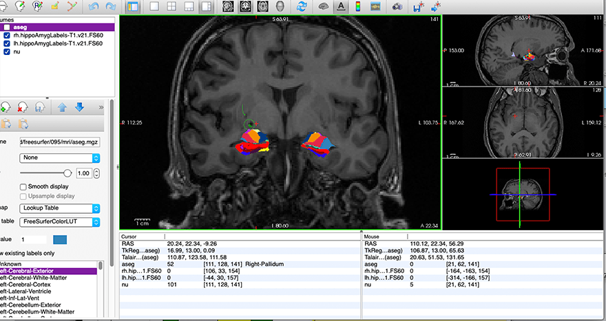
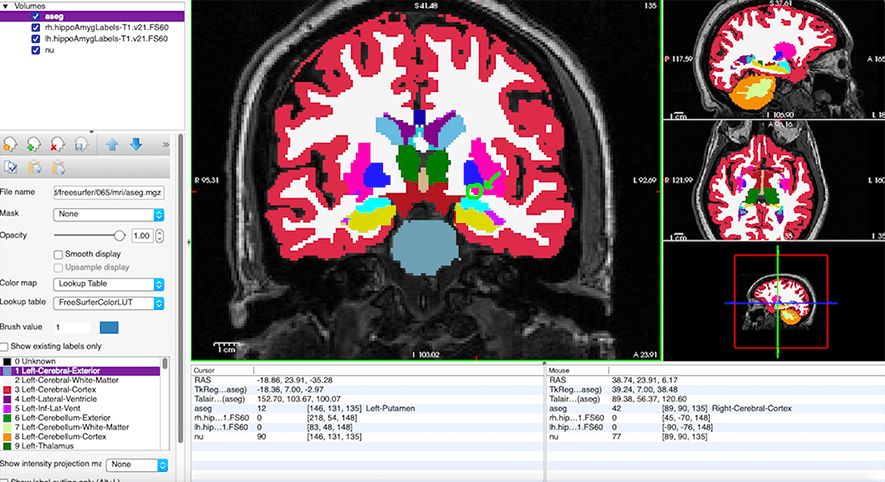
-We have come across about 5 subjects' medical nuclei masks that are located in the Putamen region (more superior/detached from the rest of the amygdala) on one side of the brain (in some cases just the right side, in other cases just the left side). In all of the other subjects, we don't see that same detached looking segmentation into the putamen on one side as these few show. I will attach screenshots to show this.
-I have checked the aseg for these subjects as suggested in the previous question, and the hippocampus, amygdala, and putamen all look fine there. I have also checked the output volumes for the medial nucleus of these subjects and the numbers all look fine.
-So, my main question is: If the placement of the medial nucleus is wrong in the segmentation (detached/in the putamen as shown) but the aseg looks correct, can we still use those values reliably?
Thank you!
Angel Hammond
Research Specialist
Center for Healthy Minds
University of Wisconsin-Madison

Dear Angel, It’s hard to tell without seeing the segmentation in 3D. Would you mind sharing, please? Cheers, /Eugenio
Juan Eugenio Iglesias Senior research fellow CMIC (UCL), MGH (HMS) and CSAIL (MIT) http://www.jeiglesias.com
From: freesurfer-bounces@nmr.mgh.harvard.edu on behalf of Angel Hammond ahammond3@wisc.edu Reply-To: Freesurfer support list freesurfer@nmr.mgh.harvard.edu Date: Monday, August 10, 2020 at 10:11 To: Freesurfer support list freesurfer@nmr.mgh.harvard.edu Subject: [Freesurfer] Reposting a Question RE: Hippocampal/Amygdala subfield segmentation - Medial Nucleus Errors
External Email - Use Caution
Hello Freesurfer Experts,
I wanted to re-post a portion of a question that a previous team member posted on June 20th, 2019 (See: Reposting a Question RE: Hippocampal/Amygdala subfield segmentation - Anterior Amygdala Area / Medial Nucleus Errors)
We have visually checked 116 subjects that have been processed using
Freesurfer's latest hippocampal/amygdala subfield segmentation
algorithm (Development version 20180518 https://surfer.nmr.mgh.harvard.edu/fswiki/HippocampalSubfieldsAndNucleiOfAmygdala)
and came across a few cases that had questionable segmentation. The T1s had were processed using
FS 5.3, and we used the command "segmentHA_T1.sh" from the latest Dev
version.
-We have come across about 5 subjects' medical nuclei masks that are located in the Putamen region (more superior/detached from the rest of the amygdala) on one side of the brain (in some cases just the right side, in other cases just the left side). In all of the other subjects, we don't see that same detached looking segmentation into the putamen on one side as these few show. I will attach screenshots to show this.
-I have checked the aseg for these subjects as suggested in the previous question, and the hippocampus, amygdala, and putamen all look fine there. I have also checked the output volumes for the medial nucleus of these subjects and the numbers all look fine.
-So, my main question is: If the placement of the medial nucleus is wrong in the segmentation (detached/in the putamen as shown) but the aseg looks correct, can we still use those values reliably?
Thank you!
Angel Hammond
Research Specialist
Center for Healthy Minds
University of Wisconsin-Madison
External Email - Use Caution
Hi Eugenio,
Happy to share that with you. I am pretty new to Freesurfer, so could you clarify what volumes you would like me to share? I currently have "aseg.mgz", "rh.hippoAmygLabels-T1.v21.FS60.mgz", "lh.hippoAmygLabels-T1.v21.FS60.mgz", and "nu.mgz" loaded in my 2d screenshots I sent. Also, would you like me to send these using the FTP file exchange?
Thank you!
Angel Hammond
Research Specialist
Center for Healthy Minds
University of Wisconsin-Madison
________________________________ From: freesurfer-bounces@nmr.mgh.harvard.edu freesurfer-bounces@nmr.mgh.harvard.edu on behalf of Iglesias Gonzalez, Juan E. JIGLESIASGONZALEZ@mgh.harvard.edu Sent: Monday, August 10, 2020 9:14 AM To: Freesurfer support list freesurfer@nmr.mgh.harvard.edu Subject: Re: [Freesurfer] Reposting a Question RE: Hippocampal/Amygdala subfield segmentation - Medial Nucleus Errors
Dear Angel,
It’s hard to tell without seeing the segmentation in 3D. Would you mind sharing, please?
Cheers,
/Eugenio
Juan Eugenio Iglesias
Senior research fellow
CMIC (UCL), MGH (HMS) and CSAIL (MIT)
From: freesurfer-bounces@nmr.mgh.harvard.edu on behalf of Angel Hammond ahammond3@wisc.edu Reply-To: Freesurfer support list freesurfer@nmr.mgh.harvard.edu Date: Monday, August 10, 2020 at 10:11 To: Freesurfer support list freesurfer@nmr.mgh.harvard.edu Subject: [Freesurfer] Reposting a Question RE: Hippocampal/Amygdala subfield segmentation - Medial Nucleus Errors
External Email - Use Caution
Hello Freesurfer Experts,
I wanted to re-post a portion of a question that a previous team member posted on June 20th, 2019 (See: Reposting a Question RE: Hippocampal/Amygdala subfield segmentation - Anterior Amygdala Area / Medial Nucleus Errors)
We have visually checked 116 subjects that have been processed using
Freesurfer's latest hippocampal/amygdala subfield segmentation
algorithm (Development version 20180518 https://surfer.nmr.mgh.harvard.edu/fswiki/HippocampalSubfieldsAndNucleiOfAmygdala)
and came across a few cases that had questionable segmentation. The T1s had were processed using
FS 5.3, and we used the command "segmentHA_T1.sh" from the latest Dev
version.
-We have come across about 5 subjects' medical nuclei masks that are located in the Putamen region (more superior/detached from the rest of the amygdala) on one side of the brain (in some cases just the right side, in other cases just the left side). In all of the other subjects, we don't see that same detached looking segmentation into the putamen on one side as these few show. I will attach screenshots to show this.
-I have checked the aseg for these subjects as suggested in the previous question, and the hippocampus, amygdala, and putamen all look fine there. I have also checked the output volumes for the medial nucleus of these subjects and the numbers all look fine.
-So, my main question is: If the placement of the medial nucleus is wrong in the segmentation (detached/in the putamen as shown) but the aseg looks correct, can we still use those values reliably?
Thank you!
Angel Hammond
Research Specialist
Center for Healthy Minds
University of Wisconsin-Madison

You can follow the instructions below (but send the email to Eugenio:).
Eugenio, you will be able to find the data in /space/incoming
From the linux command line, Create the file you want to upload, eg, cd $SUBJECTS_DIR tar cvfz subject.tar.gz ./subject Now log into our anonymous FTP site: ftp surfer.nmr.mgh.harvard.edu It will ask you for a user name: use "anonymous" (no quotes) It will ask you for a password: use "anonymous" (no quotes) cd transfer/incoming binary put subject.tar.gz Send an email that the file has been and the name of the file.
On 8/10/2020 12:42 PM, Angel Hammond wrote:
External Email - Use Caution
Hi Eugenio,
Happy to share that with you. I am pretty new to Freesurfer, so could you clarify what volumes you would like me to share? I currently have "aseg.mgz", "rh.hippoAmygLabels-T1.v21.FS60.mgz", "lh.hippoAmygLabels-T1.v21.FS60.mgz", and "nu.mgz" loaded in my 2d screenshots I sent. Also, would you like me to send these using the FTP file exchange?
Thank you!
*Angel Hammond*
*Research Specialist
Center for Healthy Minds
University of Wisconsin-Madison
*From:* freesurfer-bounces@nmr.mgh.harvard.edu freesurfer-bounces@nmr.mgh.harvard.edu on behalf of Iglesias Gonzalez, Juan E. JIGLESIASGONZALEZ@mgh.harvard.edu *Sent:* Monday, August 10, 2020 9:14 AM *To:* Freesurfer support list freesurfer@nmr.mgh.harvard.edu *Subject:* Re: [Freesurfer] Reposting a Question RE: Hippocampal/Amygdala subfield segmentation - Medial Nucleus Errors
Dear Angel,
It’s hard to tell without seeing the segmentation in 3D. Would you mind sharing, please?
Cheers,
/Eugenio
Juan Eugenio Iglesias
Senior research fellow
CMIC (UCL), MGH (HMS) and CSAIL (MIT)
*From: *freesurfer-bounces@nmr.mgh.harvard.edu on behalf of Angel Hammond ahammond3@wisc.edu *Reply-To: *Freesurfer support list freesurfer@nmr.mgh.harvard.edu *Date: *Monday, August 10, 2020 at 10:11 *To: *Freesurfer support list freesurfer@nmr.mgh.harvard.edu *Subject: *[Freesurfer] Reposting a Question RE: Hippocampal/Amygdala subfield segmentation - Medial Nucleus Errors
* External Email - Use Caution *
Hello Freesurfer Experts,
I wanted to re-post a portion of a question that a previous team member posted on June 20th, 2019 /(See: Reposting a Question RE: Hippocampal/Amygdala subfield segmentation - Anterior Amygdala Area / Medial Nucleus Errors)/
We have visually checked 116 subjects that have been processed using
Freesurfer's latest hippocampal/amygdala subfield segmentation
algorithm (Development version 20180518 https://surfer.nmr.mgh.harvard.edu/fswiki/HippocampalSubfieldsAndNucleiOfAmygdala)
and came across a few cases that had questionable segmentation. The T1s had were processed using
FS 5.3, and we used the command "segmentHA_T1.sh" from the latest Dev
version.
-We have come across about 5 subjects' medical nuclei masks that are located in the Putamen region (more superior/detached from the rest of the amygdala) on one side of the brain (in some cases just the right side, in other cases just the left side). In all of the other subjects, we don't see that same detached looking segmentation into the putamen on one side as these few show. I will attach screenshots to show this.
-I have checked the aseg for these subjects as suggested in the previous question, and the hippocampus, amygdala, and putamen all look fine there. I have also checked the output volumes for the medial nucleus of these subjects and the numbers all look fine.
*-So, my main question is:* If the placement of the medial nucleus is wrong in the segmentation (detached/in the putamen as shown) but the aseg looks correct, can we still use those values reliably?
Thank you!
*Angel Hammond*
Research Specialist
Center for Healthy Minds
University of Wisconsin-Madison
Freesurfer mailing list Freesurfer@nmr.mgh.harvard.edu https://mail.nmr.mgh.harvard.edu/mailman/listinfo/freesurfer
External Email - Use Caution
Hi Eugenio,
I have uploaded the files to the FTP under the file named 065_help.zip. Please let me know if there are any issues accessing them.
Thank you!
Angel Hammond
Research Specialist
Center for Healthy Minds
University of Wisconsin-Madison
________________________________ From: freesurfer-bounces@nmr.mgh.harvard.edu freesurfer-bounces@nmr.mgh.harvard.edu on behalf of Iglesias Gonzalez, Juan E. JIGLESIASGONZALEZ@mgh.harvard.edu Sent: Monday, August 10, 2020 9:14 AM To: Freesurfer support list freesurfer@nmr.mgh.harvard.edu Subject: Re: [Freesurfer] Reposting a Question RE: Hippocampal/Amygdala subfield segmentation - Medial Nucleus Errors
Dear Angel,
It’s hard to tell without seeing the segmentation in 3D. Would you mind sharing, please?
Cheers,
/Eugenio
Juan Eugenio Iglesias
Senior research fellow
CMIC (UCL), MGH (HMS) and CSAIL (MIT)
From: freesurfer-bounces@nmr.mgh.harvard.edu on behalf of Angel Hammond ahammond3@wisc.edu Reply-To: Freesurfer support list freesurfer@nmr.mgh.harvard.edu Date: Monday, August 10, 2020 at 10:11 To: Freesurfer support list freesurfer@nmr.mgh.harvard.edu Subject: [Freesurfer] Reposting a Question RE: Hippocampal/Amygdala subfield segmentation - Medial Nucleus Errors
External Email - Use Caution
Hello Freesurfer Experts,
I wanted to re-post a portion of a question that a previous team member posted on June 20th, 2019 (See: Reposting a Question RE: Hippocampal/Amygdala subfield segmentation - Anterior Amygdala Area / Medial Nucleus Errors)
We have visually checked 116 subjects that have been processed using
Freesurfer's latest hippocampal/amygdala subfield segmentation
algorithm (Development version 20180518 https://surfer.nmr.mgh.harvard.edu/fswiki/HippocampalSubfieldsAndNucleiOfAmygdala)
and came across a few cases that had questionable segmentation. The T1s had were processed using
FS 5.3, and we used the command "segmentHA_T1.sh" from the latest Dev
version.
-We have come across about 5 subjects' medical nuclei masks that are located in the Putamen region (more superior/detached from the rest of the amygdala) on one side of the brain (in some cases just the right side, in other cases just the left side). In all of the other subjects, we don't see that same detached looking segmentation into the putamen on one side as these few show. I will attach screenshots to show this.
-I have checked the aseg for these subjects as suggested in the previous question, and the hippocampus, amygdala, and putamen all look fine there. I have also checked the output volumes for the medial nucleus of these subjects and the numbers all look fine.
-So, my main question is: If the placement of the medial nucleus is wrong in the segmentation (detached/in the putamen as shown) but the aseg looks correct, can we still use those values reliably?
Thank you!
Angel Hammond
Research Specialist
Center for Healthy Minds
University of Wisconsin-Madison
freesurfer@nmr.mgh.harvard.edu
-
 Angel Hammond
 -
 Douglas N. Greve
 -
 Iglesias Gonzalez, Juan E.