
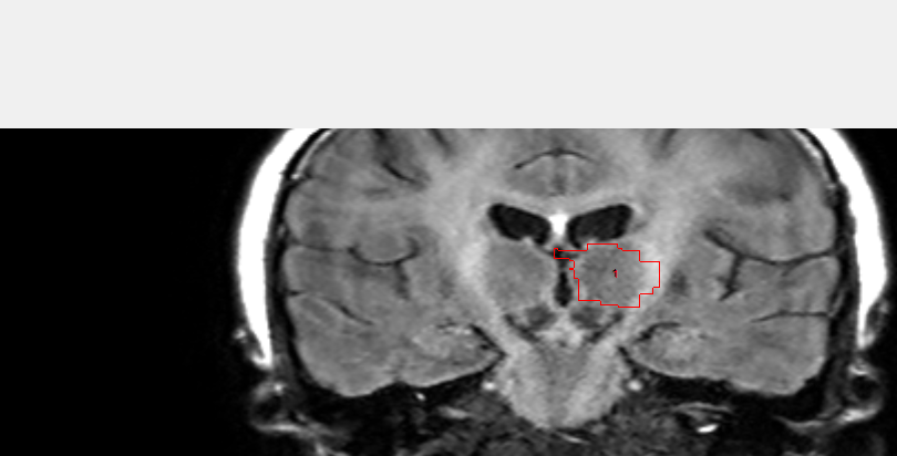
I have attached a screenshot of one of the slices. from what I see it is going a little far in the medial and lateral parts. I am using a 3TC image taken by Philips device. Is there any way to limit the region of interest before running the recon-all to only segment the thalamus and may give better results?
Thank you
Dorsa
On Wed, Oct 19, 2016 at 5:21 PM, Bruce Fischl fischl@nmr.mgh.harvard.edu wrote:
Hi Dorsa
why do you think it is going out too far? What intensity volume are you overlaying it on? Many segmentations miss the lateral parts of the thalamus since they have so much myelin in them
cheers Bruce On Wed, 19 Oct 2016, Dorsa Haji Ghaffari wrote:
Hi, I have done the sub-cortical segmentation in Freesurfer and have
extracted the left thalamus, when I put
it on top of the original MRI, it seems like it is getting out of the
border of thalamus, also it is not
smooth. I wanted to ask if anyone knows how to limit the computation
only to thalamus, and also make it
trace the thalamus more accurately?
Thank you
Dorsa
Freesurfer mailing list Freesurfer@nmr.mgh.harvard.edu https://mail.nmr.mgh.harvard.edu/mailman/listinfo/freesurfer
The information in this e-mail is intended only for the person to whom it is addressed. If you believe this e-mail was sent to you in error and the e-mail contains patient information, please contact the Partners Compliance HelpLine at http://www.partners.org/complianceline . If the e-mail was sent to you in error but does not contain patient information, please contact the sender and properly dispose of the e-mail.